Supracerebellar Transventricular Approach
This is a preview. Check to see if you have access to the full video. Check access
Resection of a Posterior Third Ventricular Tumor Via the Supracerebellar Approach
General Considerations
Small tumors in the posterior third ventricle, such as cavernous malformations, pose a significant technical challenge during their resection. Because of their small size, they do not significantly displace deep structures and therefore cannot create a suitable supratentorial operative corridor.
In these situations, I have attempted to use the supracerebellar transventricular approach. The small size of these tumors may expand and affect the tectum, and provides just enough space for me to work through the attenuated nervous tissue. Gaze disturbances are a risk, but most deficits are temporary.
The specific region of interest is the posterior segment of the third ventricle. The aqueduct and the suprapineal recess demarcate this segment.
Figure 1: Small, very posterior third ventricular tumors expand an opening around the pineal gland and within the tectum that allows their exposure via the supracerebellar transventricular approach.
Diagnosis and Evaluation
For a general discussion of diagnosis and evaluation for ventricular tumors, see the Principles of Intraventricular Surgery chapter.
Because of their proximity to the third ventricle and the aqueduct, patients with tumors in this region often present with obstructive hydrocephalus. Parinaud’s syndrome may develop with intratumoral hemorrhage (pineal apoplexy). The tumors in this area that demand resection despite their small size include cavernous malformations, pineoblastomas, and pineal parenchymal tumors of intermediate differentiation.
Figure 2: A posterior third ventricular pineal parenchymal tumor of intermediate differentiation is shown (upper row). This tumor was removed via the supracerebellar transventricular approach (lower row)(please see Figure 8 and the video at the beginning of this chapter.)
Indications for Surgery
Because of the very limited exposure afforded via the trans-pineal or trans-tectal corridor, large and especially vascular tumors are not ideal candidates. Hydrocephalus and uncertainty regarding diagnosis are reasonable indications for surgical intervention. Cavernous malformations that have caused repetitive symptomatic hemorrhages also require excision.
Preoperative Considerations
A careful study of the preoperative magnetic resonance images should determine the location of the internal cerebral veins and the veins of Rosenthal and Galen, in relation to the tumor. If the veins are displaced posteriorly and ventrally rather than superiorly, the supracerebellar transventricular approach is contraindicated.
The use of an external ventricular drain in the presence of hydrocephalus is advised.
Operative Anatomy
The relevant anatomy is discussed in the Midline Supracerebellar Craniotomy and Anatomy of the Ventricular System chapters.
Click here to view the interactive module and related content for this image.
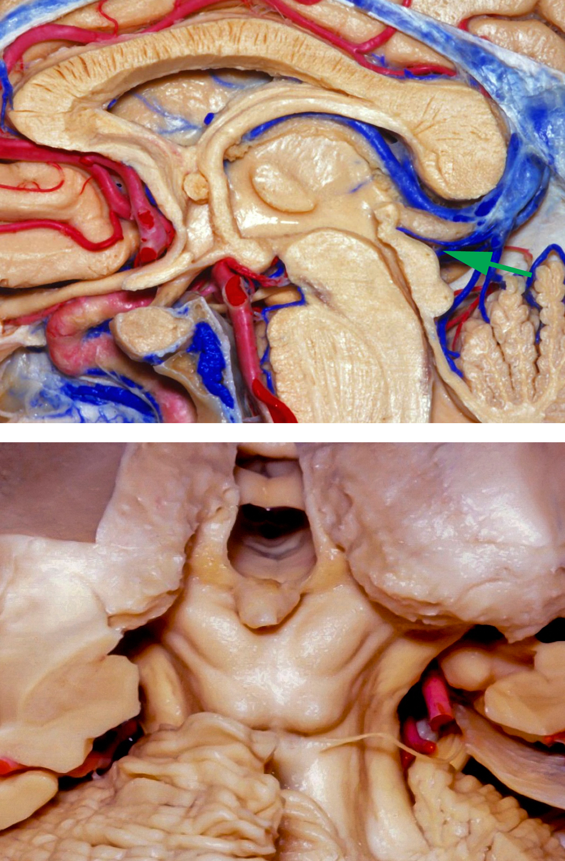
Figure 3: The operative route of the supracerebellar transventricular trajectory is demonstrated using the green arrow (upper image). A view of the ventricle through the suprapineal trajectory is shown (lower image). This approach is suitable when the posterior ventricular lesion has expanded the posterior wall of the ventricle either above or more commonly below the pineal gland and eroded through the tectum (images courtesy of Rhoton, AL).
Click here to view the interactive module and related content for this image.
Figure 4: The midline (left image) and left paramedian (right image) supracerebellar trajectories allow entry into the third ventricle based on the route created by the tumor. Note how the anatomy of the veins will dictate the feasibility of this approach (images courtesy of Rhoton, AL).
SUPRACEREBELLAR TRANSVENTRICULAR APPROACH
The infratentorial supracerebellar approach is favored for small posterior third ventricular lesions such as cavernous malformations, even if the lesion is not primarily within the pineal region. The advantages of this approach are substantial and include a minimal risk of injury to the adjacent structures and avoidance of supratentorial contents, including conservation of the corpus callosum. In my experience, the posterior pulvinar can be gently manipulated without significant risks.
The disadvantages of this approach include the vulnerability of the habenula, vein of Galen, and quadrigeminal plate. Most deficits related to manipulation of the tumor-affected tectum are temporary.
The technical details of the midline supracerebellar and paramedian supracerebellar craniotomies are discussed elsewhere in this Atlas. Although the unilateral paramedian craniotomy is adequate, most neurosurgeons prefer to use the midline supracerebellar approach to reach such deep-seated midline masses. Therefore, the following discussion refers to this type of exposure.
INTRADURAL PROCEDURE
A standard midline suboccipital supracerebellar craniotomy is performed.
Figure 5: The midline supracerebellar approach is used to expose the tectum. A posterior third ventricular tumor usually attenuates or affects the superior tectum and displaces the posterior pulvinar. First, I identify the position of the diencephalic veins, including the vein of Galen; its posterior displacement will adversely affect the resection corridor and place the vein between the surgeon and the tumor. The tumor may also engulf these veins, and the operator may inadvertently injure them during the early stages of the operation.
Figure 6: The transtectal exposure is limited. In these cases, ring curettes may be used to debulk the tumor from within the ventricle.
Figure 7: At the end of resection, a view of the internal cerebral veins and the anterior third ventricle is evident. The blind spots are numerous and include the anterior edges of the tectum, just anterior to the “lips” of the resection cavity. The use of angled endoscopes or mirrors is highly recommended. Blind pulling or traction on the tumor can place the walls of the ventricle or associated veins at risk.
Figure 8: The tumor in Figure 2 is exposed via the midline supracerebellar approach with the patient in the sitting position. Note the discoloration of the left tectum (left image, at the tip of suction device). Removal of the affected tectum allowed entry into the ventricle and gross total resection of the mass (right photo).
Closure and Postoperative Considerations
For a detailed discussion of recommendations for closure and postoperative care of patients with ventricular tumors, please see the Principles of Intraventricular Surgery chapter.
Pearls and Pitfalls
- The described approach is applicable in very select cases. The presence of diencephalic veins and intact tectum significantly limits its utility. Special anatomical considerations are necessary to justify its use.
Please login to post a comment.


