Deep Subcortical AVMs
This is a preview. Check to see if you have access to the full video. Check access
Microsurgical Resection of a Superior Thalamic AVM
Please note the relevant information for patients suffering from arteriovenous malformations is presented in another chapter. Please click here for patient-related content.
Subcortical arteriovenous malformations (AVMs) are formidable pathologies given their deep, highly eloquent parenchymal locations. Advanced knowledge of the microsurgical anatomy of the core hemispheric structures and their indispensable vascular supply is essential for safe and complete resection of these deep and insular AVMs.
Many of these malformations should be exposed during surgery only if the nidus is compact and reaches a safe ependymal or pial surface, if the patient has suffered a hemorrhagic event resulting in neurologic deficits, or if radiosurgery treatment has failed. Appropriate patient selection is imperative for acceptable results.
Operative Anatomy
The hemispheric core or basal nuclear structures lie medial and deep to the insula and act as a central relay station. They are primarily composed of the caudate nucleus, putamen, globus pallidus (collectively referred to as the basal ganglia), and the thalamus. The internal capsule is the major white matter tract between these core hemispheric components. The insula acts as the outermost wall of the basal ganglia and is often called the fifth lobe.
Small arterial perforators from the circle of Willis nourish the deep parenchymal territories. These angiographically invisible small end-arteries carry minimal to no collateral support and are vital to life. Some of these white matter feeders contribute to arteriovenous shunting and are difficult to identify and control when encountered during circumdissection of an AVM. Many of these vessels are faced late in dissection because the AVM is located between the surgeon and these feeders. Therefore, a thorough anatomic study of these perforating vessels is essential.
The perforating arteries of the anterior circulation include:
- Medial lenticulostriate arteries are typically eight perforators that arise from the A1 segment of the anterior cerebral artery (ACA) or the proximal M1 and supply the suprachiasmatic hypothalamus, third ventricle, dorsal optic chiasm, nerve, and tract.
- Recurrent artery of Heubner is the most medial perforator among the medial lenticulostriate arteries and arises from the A1-A2 junction (commonly from the proximal A2) segment. It courses along with the rest of the medial lenticulostriate arteries to supply the head of caudate, adjacent internal capsule, anterior putamen, and globus pallidus. It courses parallel, but in the opposite direction to the A1.
- Lateral/intermediate lenticulostriate arteries are commonly ten perforators that arise from the distal M1 segment of the middle cerebral artery (MCA) and supply the internal capsule, globus pallidus, and head/body of the caudate nucleus. In patients with an early MCA bifurcation, these arteries may arise from the proximal M2 trunks.
- Anterior thalamoperforators are generally eight perforators which arise from the supralateral surfaces of the posterior communicating artery (PCoA) and supply the ventral thalamus, posterior hypothalamus, anterior third of the optic tract, posterior limb of the internal capsule, and subthalamic nucleus.
- Insular perforators are not conspicuous in a normal brain, but they become prominent in the presence of arteriovenous shunting of an AVM in the Sylvian, insular, or lateral basal ganglia regions. These perforating vessels arise from the M2 segments of the MCA.
The perforating arteries of the posterior circulation include:
- Posterior thalamoperforators are commonly four perforators that arise from the P1 segment of the posterior cerebral artery (PCA). When only a single perforator is present, it is referred to as the artery of Percheron. These vessels supply the thalamus, posterior hypothalamus, subthalamus, and medial midbrain.
- Peduncular perforators arise from the P2 segment of the PCA and supply the corticospinal/corticobulbar tracts, substantia nigra, red nucleus, and midbrain tegmentum.
- Circumflex perforators arise from P1 and P2 segments of the PCA and go around the midbrain (hence they are described as circumflex). Short circumflex perforators supply the geniculate bodies, and long circumflex perforators supply the quadrigeminal plate.
- Thalamogeniculate arteries are usually two or three vessels that arise from the P2 segment of the PCA and supply the posterior half of the lateral thalamus, the posterior limb of the internal capsule, and the posterior optic tract.
Figure 1: The origins of the perforating arteries, mentioned above, from an inferior view of the circle of Willis are illustrated (images courtesy of AL Rhoton, Jr).
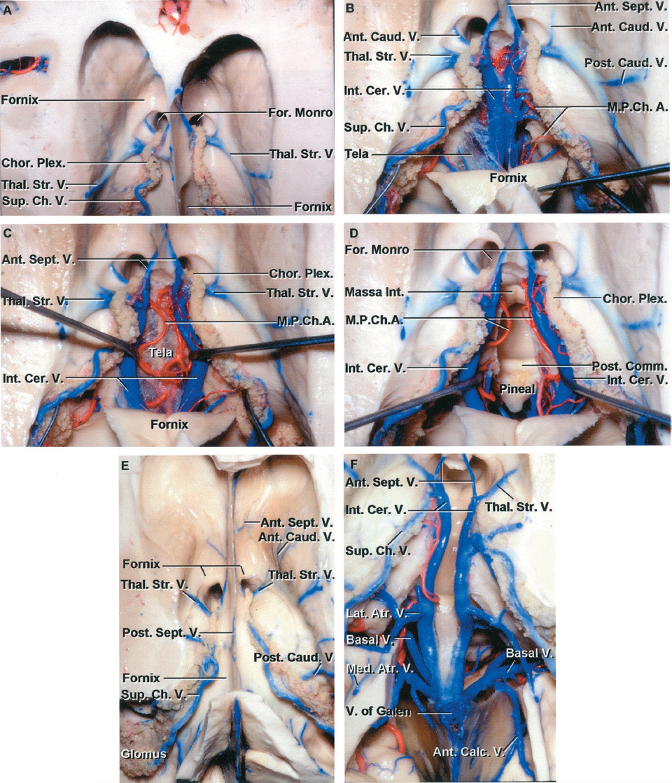
The major venous drainage patterns of the deep core nuclei include:
- Anterior and posterior caudate veins drain the caudate nucleus and terminate in the thalamostriate vein.
- Internal cerebral veins (ICVs) are paired veins housed in the velum interpositum in the roof of the third ventricle.
- Vein of Galen receives the flow from the ICVs, the basal veins of Rosenthal, and the precentral cerebellar vein. I subsequently joins the straight sinus.
- Lateral atrial vein drains the lateral wall of the atrium, posterior thalamus, and the body of caudate into the basal vein of Rosenthal.
- Superficial and deep Sylvian veins drain the insular region via their anastomoses with the basal vein of Rosenthal.
Click here to view the interactive module and related content for this image.
Figure 2: The venous anatomy for drainage of the central core of the hemisphere is illustrated (images courtesy of AL Rhoton, Jr).
SUBCORTICAL/DEEP/INSULAR ARTERIOVENOUS MALFORMATION RESECTION
Basal Ganglia AVMs
Basal ganglia AVMs are found deep to the insula and superficial to the posterior limb of the internal capsule. Putaminal and caudate AVMs are more accessible and therefore operable compared with their deeper counterparts (globus pallidus AVMs).
Putaminal AVMs
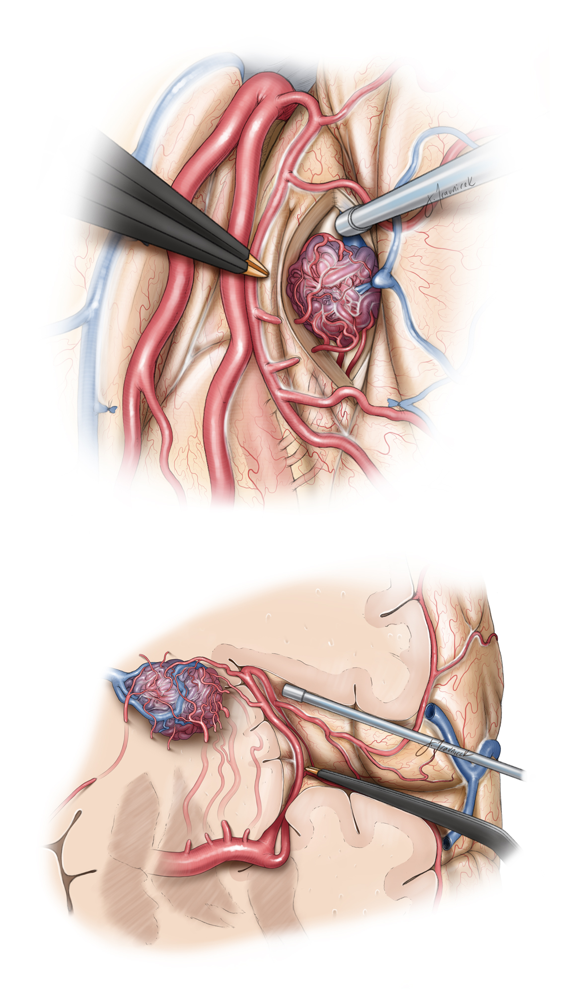
The optimal operative trajectory to putaminal AVMs includes the use of a standard pterional craniotomy and the transsylvian-transinsular approach. This approach requires an extensive Sylvian fissure split to grant permissive exposure of the nidus. The fissure dissection is discussed in the chapter titled Techniques of Sylvian Fissure Split.
The draining veins, including the caudate, thalamostriate veins, and the basal vein of Rosenthal, are uncovered only at the medial limit of the dissection; therefore, they do not serve as a roadmap to guide AVM dissection. The arterial supply to the lateral basal ganglia AVMs derives from the insular perforators laterally and the lateral/medial lenticulostriate arteries inferiorly.
The insular perforators are encountered en route to the medial pole of the AVM and are disrupted using microclips or, if readily amenable, bipolar electrocautery. Subsequent change in the color of the main draining vein (the deep Sylvian vein) serves to confirm complete occlusion of the arterial feeders.
The most important aspect of this dissection is to differentiate non-AVM perforating vessels from the AVM-associated ones. Indiscriminate coagulation can harm bystander vessels.
Figure 3: Please note the surgeon's view of the operative pathoanatomy for a left-sided basal ganglia AVM via the transsylvian-transinsular approach (top sketch). Note the medially projecting deep draining vein traveling away from the surgeon (coronal view, bottom sketch).
Caudate AVMs
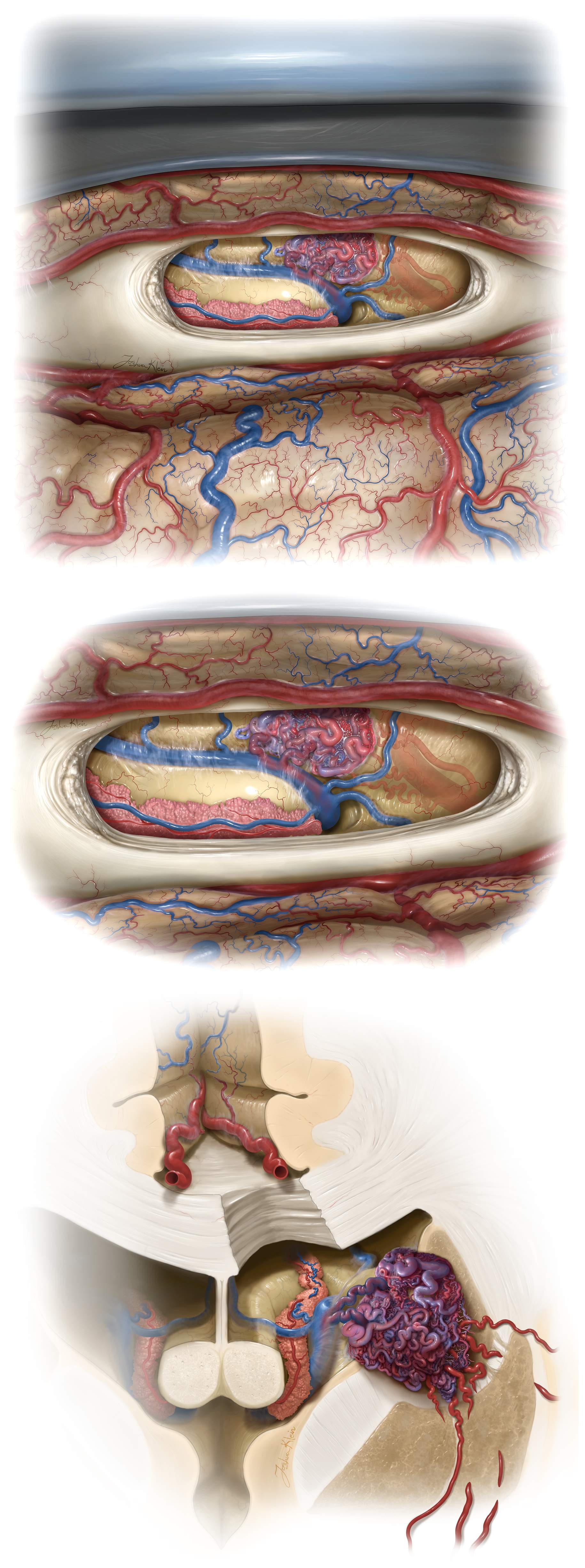
The optimal operative approach to these lesions includes the use of the contralateral transcallosal route, the same route used to access callosal AVMs discussed in the Periventricular AVMs chapter. The contralateral transcallosal approach is ideal for surgical targets that extend to the medial basal ganglia.
Figure 4: The contralateral transcallosal trajectory requires an operative lobby involving the contralateral interhemispheric fissure, supplanted by an oblique transcallosal section and expanded lateral reach via the ipsilateral ventricle. The AVM usually approaches the lateral wall of the ventricle.
The caudate and thalamostriate veins are the main draining veins and serve as early landmarks through this operative exposure. The medial lenticulostriates and recurrent artery of Heubner are the main arterial feeders and lie along the inferomedial aspect of the dissection planes. The lateral plane is developed to identify the remaining feeders, occlude the venous drainage, and excise the nidus.
Caudate AVMs are located in highly eloquent parenchyma, and surgical resection poses significant risks if the striatal components are transgressed.
Figure 5: Note the right-sided interhemispheric transcallosal route to this left caudate AVM and the associated draining veins joining the thalamostriate vein. The lenticulostriate arteries and recurrent artery of Heubner are ghosted in to demonstrate the relevant anatomy (Top and middle.) The last illustration focuses on the anatomy at the coronal plane.
Thalamic AVMs
Thalamic AVMs that are amenable to operative intervention are divided into two major groups: superior and medial lesions. The vascular anatomy is similar between these two divisions of the thalamic AVMs.
They receive their arterial supply through the anterior thalamoperforators (arising from the PCoA), posterior thalamoperforators (emerging from the P1), and lateral/medial posterior choroidal arteries. Venous drainage is routed to the ICVs superiorly and into the basal vein of Rosenthal medially.
| Indication | Surgical Approach |
| Anterior thalamic AVM | Anterior transsylvian transinsular |
| Posterior transsylvian transinsular | |
| Superior thalamic AVM | Anterior ipsilateral transcallosal |
| Anterior contralateral transcallosal | |
| Posterior ipsilateral transcallosal | |
| Medial thalamic AVM | Transcallosal expanded transforaminal transvenous/transchoroidal |
| Posterior thalamic AVM | Posterior ipsilateral transcallosal |
| Adjacent hematoma or encephalomalacia | Transcortical (frontal) |
| Transcortical (parietal) | |
| Transcortical (temporal) |
The superior and medial thalamic lesions are most amendable and accessible to resection as they approach the ependymal surface within the third ventricle and therefore minimize the need for transgression of the thalamus. The other subtypes of thalamic AVMs can be safely exposed during surgery if they have ruptured and are therefore are accessible via nonanatomic pathways. Otherwise, radiosurgery or observation is the more reasonable treatment modality.
Superior lesions
Several approaches have been proposed to access the superior thalamus, as listed in Table 1; however, the transventricular approaches are favored because of their minimally disruptive features.
The ipsilateral transcallosal approach (please refer to the Interhemispheric Craniotomy chapter) is favored for midline lesions. I prefer the contralateral “cross court” route for lesions extending beyond the midline and centered more laterally.
The medial and lateral posterior choroidal arteries and posterior thalamoperforators are the main feeders for these lesions. These feeding vessels are first occluded following identification of their terminal vessels at the nidus along the superior thalamus. The draining veins entering the choroidal fissure are identified medially as they join the ICVs and are occluded after circumferential nidal disconnection.
Circumdissection of the AVM with occlusion of the arterial feeders is performed sequentially on the anterior, posterior, and lateral surfaces of the nidus. After mobilization of the nidus, I expose the underlying inferior arterial feeders. Next, these vessels are occluded to complete a thorough circumdissection, allowing removal of the nidus.
Figure 6: The anatomy of a superior thalamic AVM through a right-sided interhemispheric transcallosal route is demonstrated. Based on the surgeon's view (top), the choroidal feeders along the margin of the malformation are disconnected circumferentially. The coronal view (bottom) elucidates the route the the draining vein.
Figure 7: A ruptured superior thalamic AVM that resisted occlusion via radiosurgery is included. Note the location of the AVM and the feeding vessels involving the anterior and posterior thalamoperforators, and lateral/medial posterior choroidal arteries. Venous drainage is routed to the ICVs superiorly and into the basal vein of Rosenthal medially. A contralateral posterior frontal interhemispheric transfalcine transventricular approach (third row) was deemed appropriate for exposure of this lateral thalamic AVM (fourth row). This lesion was circumferentially disconnected and the choroidal vessels feeding the AVM sacrificed; the disconnected nidus and its draining vein are shown (the photos of the last row).
Medial lesions
With medially located lesions, the ipsilateral transcallosal expanded transforaminal transvenous transchoroidal approach uncovers the medial wall of the third ventricle. The main feeding anterior/posterior thalamoperforators are located at the inferior margin of dissection; this configuration adds to the risk of resection in these lesions. The nidus must be mobilized medially into the third ventricle for me to access these deep-seated feeding vessels that frequently require clipping for their control.
A similar approach must be considered when occluding the venous outflow because the basal vein of Rosenthal will be visualized only after near complete circumdissection of the nidus. The internal capsule is in close proximity, lateral to the foramen of Monro, whereas the columns of the fornix are anteromedial to the foramen. The vicinity of these vital structures requires meticulous manipulation of the adjacent neural tissues.
Thalamic lesions are treacherous, difficult to access, and located in a highly eloquent region. Resection should be attempted only if the lesion is small and the nidus is compact and reaches the ependymal surface. A previous history of hemorrhage and preexisting neurologic deficits will especially justify an operative treatment strategy.
Figure 8: Unlike caudate lesions that can benefit from the contralateral transcallosal approach, the deeper seated third ventricular lesions benefit from the ipsilateral route (top two images.) The ipsilateral hemisphere is placed in dependent position to use gravity retraction. Transection of the septal vein and anterior choroidal dissection (transcallosal expanded transforaminal transvenous transchoroidal approach) will provide ample space into the third ventricle (bottom two images.)
Figure 9: An AVM on the medial wall of the third ventricle is demonstrated. Note the large thalamoperforating feeding vessels and the draining vein joining the vein of Galen (upper rows). An anterior ipsilateral interhemispheric transchoroidal approach was used (middle images). In the left lower photo, the nidus is evident and the location of the thalamostriate vein is marked with an arrow. The right lower photo demonstrates the wall of the third ventricle following removal of the nidus.
I unfortunately caused an intraoperative rupture of a thalamic AVM by staying too close to the nidus during my compulsive attempts to preserve as much normal tissue as possible. Although preservation of the normal thalamic tissue is imperative, overzealously close dissection to the nidus can lead to an inadvertent nidal entry, causing torrential bleeding and the need for an extremely dangerous lifesaving “commando” operation within very narrow operative corridors to control bleeding while removing the deep AVM.
A Medial Thalamic AVM and the "Commando Operation"
Sylvian Cistern AVMs
Sylvian cistern AVMs reside primarily within the Sylvian fissure and the corresponding subarachnoid space (Sylvian cistern) with no significant lobar or parenchymal invasion. This feature allows dissection and resection without pial violation.
As expected, the pterional craniotomy and the transsylvian trajectory is the workhorse approach for these lesions. The patient is positioned supine and the patient’s head is turned 20 to 30 degrees contralaterally and extended 20 degrees. This head configuration places the zygoma/malar eminence as the highest point on the operative field, allowing the frontal lobe to retract under gravity.
The Sylvian fissure is split using standard microsurgical techniques. The Sylvian AVMs receive their blood supply from the M2 and M3 branches that are located deep and medial to the nidus. Venous drainage occurs via the superficial and deep Sylvian veins.
The superficial Sylvian vein is encountered during the initial steps of the operation and mobilized with the temporal lobe. The M1 is identified early through dissection of the sphenoidal section of the fissure. Distal dissection along the M1 will expose the M2/M3 feeders, which should be transected only if they have been followed distally to the nidus so that their identities as terminal feeding vessels (versus bystander or transit arteries) have been unquestionably confirmed.
The large-caliber inferomedial feeders require microclips rather than cauterization for hemostasis. En passage vessels are preserved to maintain parenchymal perfusion. The dissection proceeds while the surgeon maintains pial and parenchymal integrity until the nidus is completely mobilized and arterial feeders are thoroughly and circumferentially disrupted.
Indiscriminate coagulation in the face of bleeding is a recipe for disaster. Untangling the spaghetti of vessels within the Sylvian cisterns and identifying the bystander and transit arteries are imperative for avoiding complications.
Figure 10: The Sylvian AVMs typically do not invade the pia and are limited to the Sylvian cisterns. The M2 branches are skeletonized toward the malformation and only the AVM-associated vessels are sacrificed. Indiscriminate use of electrocoagulation within the crowded Sylvian cisterns, full of arterial branches, is prohibited in the face of bleeding.
Figure 11: The partially arterialized superficial Sylvian draining vein is encountered during the initial steps of the operation and mobilized with the temporal lobe. The M1 is identified early through dissection of the sphenoidal section of the fissure. Distal dissection along the M1 will expose the M2/M3 feeders, which should be transected only if they have been followed distally to the nidus so that their identities as terminal feeding vessels (versus bystander or transit arteries) have been confirmed.
Insular AVMs
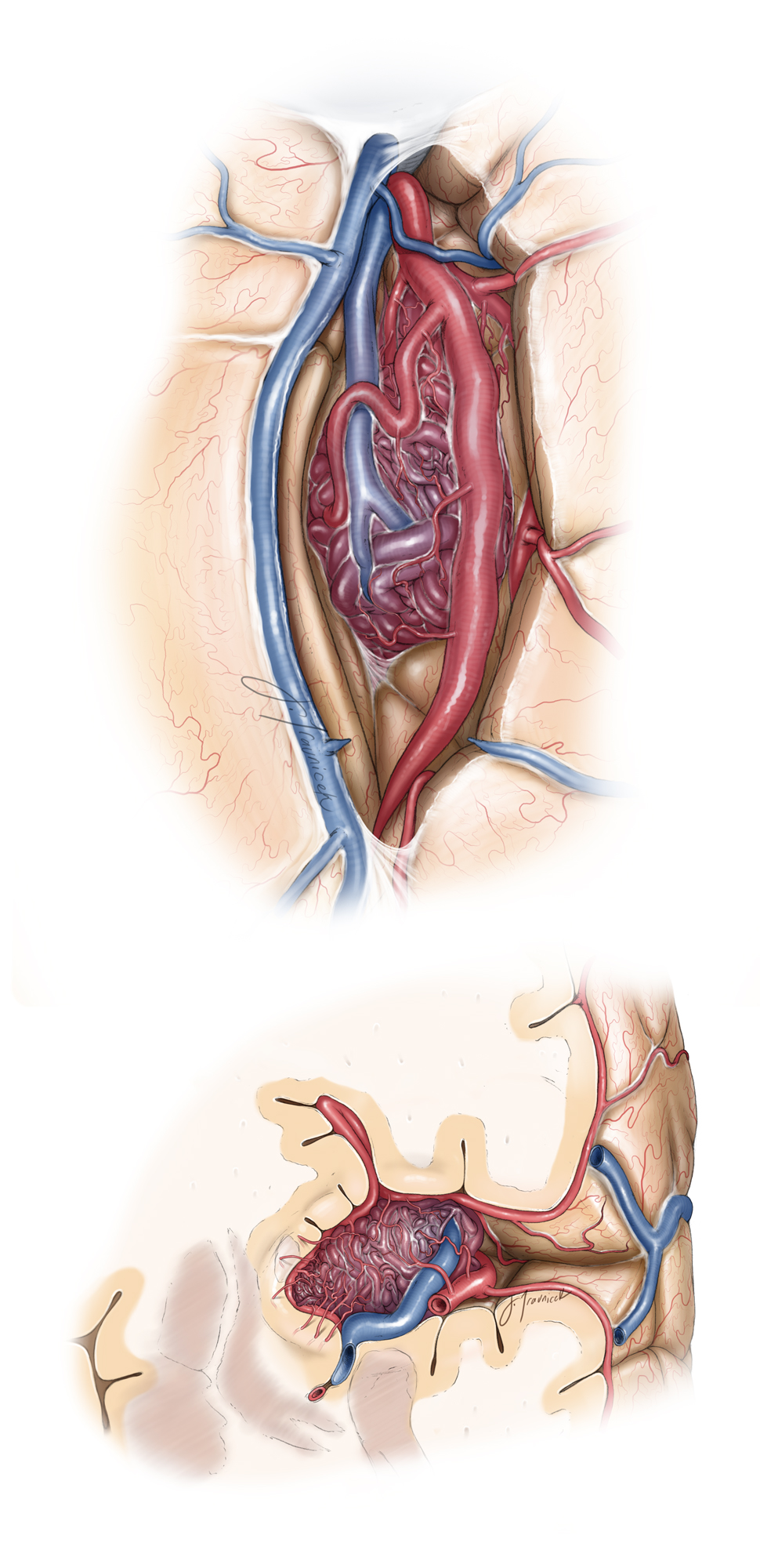
Insular AVMs are found lateral to the basal ganglia and the claustrum, but they affect the limen insula. The approach to this type of AVM is similar to that for Sylvian AVMs. After a generous Sylvian fissure split, the arterial supply from the M2 segments lateral and rostral to the nidus are disconnected.
The superficial and deep Sylvian veins are the predominant draining vessels. The superficial arteries are pursued via their skeletonization deep into the fissure and to the level of the MCA trunks; the smaller insular feeders over the insula are disconnected early. The M1 segment is isolated, and the deep lateral lenticulostriate feeders are found, but left intact until the AVM nidus is more clearly defined.
Working channels between the M2 and M3 branches allow pial transgression into the insula while the surgeon follows the arterialized vein. Next, the nidus is circumdissected. Afterwards, I mobilize the nidus to inspect its inferior and medial margins so that I can clearly identify its feeders from the lenticulostriates and its draining veins into the caudate vein.
Once the deep Sylvian vein turns dark blue, it confirms the disruption of all major arterial feeders. Only then may the other veins be occluded and the nidus excised. Despite being located in a relatively noneloquent cortex, dissection in the dominant hemisphere should be performed with care to prevent damage to the speech areas of the brain.
Once again, the main source of morbidity from this operation is collateral damage to the non-AVM perforating vessels.
Figure 12: The pathoanatomy of a typical insular AVM on the left-side is demonstrated. Note the M2 feeding arteries (surgeon's view, top sketch). The coronal image (bottom sketch) illustrates the route of the draining vein and emphasizes the contribution of the deep feeding lentriculostriate arterioles.
Contributor: Farhan A. Mirza, MBBS
References
Lawton MT. Seven AVMs: Tenets and Techniques for Resection. New York, Stuttgart: Thieme Medical Publishers, 2014.
Spetzler RF. Comprehensive Management of Arteriovenous Malformations of the Brain and Spine. Cambridge University Press, 2015.
Please login to post a comment.


