Spinal Cord Cavernous Malformation
Please note the relevant information for patients suffering from cavernous malformations is presented in another chapter. Please click here for patient-related content.
Schultze performed the first successful resection of a spinal cavernous malformation (CM) in 1912. With improvements in imaging, spinal CMs have been included in the differential diagnosis of patients presenting with various spinal cord pathology.
Spinal cord CMs account for approximately 5% of CMs of the central nervous system. They most frequently present in the thoracic spinal cord. The goal of treatment is complete resection, which can be achieved with a low morbidity rate in the majority of patients.
Clinical Manifestations and Diagnosis
Similar to brainstem and deep CMs, spinal CMs are more likely to present symptomatically than cerebral CMs. The clinical symptoms and their time course depend on the location of the CM in the spinal cord, the progression rate of the lesion, and presence or absence of hemorrhage.
Acute neurologic deficits occur with a recent hemorrhage. Patients may suffer from acute motor or sensory deficits, bowel and bladder dysfunction, and pain. Chronic myelopathy may result from an enlarging lesion or repeated small microhemorrhages.
As the associated hemoglobin breaks down, toxic metabolites are released, causing destruction and irritation of the spinal cord tracts. Episodic symptoms may occur with small hemorrhages that resolve as the blood products are reabsorbed. This episodic pattern may be misdiagnosed as transverse myelitis or multiple sclerosis.
Magnetic resonance imaging (MRI) is the modality of choice for diagnosis of CMs. Angiography is of minimal value unless an arteriovenous malformation needs to be ruled out. Patients with a family history of CMs should also undergo imaging for intracranial lesions. Up to 20% of the patients with spinal CMs also harbor intracranial lesions.
Figure 1: A conus medullaris CM is shown. This lesion led to paraparesis and subsequently was resected.
Surgical Indications and Planning
As with all CMs, the risks and benefits of surgery must be weighed against the natural history of the lesion. Annual hemorrhage rates can range from 1% to 7%, with a recent metaanalysis study suggesting an estimated annual rate of 2.1%.
Patients who present with symptomatic lesions should be considered for treatment. Resection of a lesion within 3 months of the occurrence of symptoms may correlate positively with a favorable neurologic outcome. Patients with transient minor symptoms or asymptomatic lesions may not need a resective strategy. However, if the lesion abuts the pial surface and is associated with transient symptoms, the lesion may be considered for resection if the surgical risk is considered relatively minimal.
I reimage asymptomatic lesions only if new symptoms develop because lack of new symptoms most likely will not change the management strategy. As discussed in the brainstem CM chapter, the two-point method is used on a T1-weighted image to plan the shortest operative trajectory to the lesion. The shortest route may not always be the best one; a longer route to the lesion should be chosen if it will avoid the eloquent tracts.
MICROSURGICAL RESECTION OF SPINAL CORD CAVERNOUS MALFORMATIONS
Somatosensory and motor evoked potentials monitoring is required for all spinal cord CM cases to warn the surgeon regarding potential risky maneuvers. This methodology should not endow the surgeon with a false sense of security.
On the other hand, although the monitoring technology provides important feedback about altering dynamic retraction maneuvers, it should not prevent the surgeon from removing well-defined non-infiltrative lesions such as CMs.
The majority of spinal cord CMs are located or point dorsally; thus, a posterior, posterolateral, or lateral approach can reach the pial surface where the CM is presenting itself. A laminoplasty or laminectomy at the level of the lesion should provide a reasonable visualization of the lesion with its typical bluish/purple pial discoloration.
Lateral lesions may be adequately exposed via a hemilaminectomy, facetectomy and partial pediculectomy, although a total laminectomy is necessary for lesions crossing the midline. For deeper dorsal lesions, Doppler ultrasonography may be used to precisely locate the lesion and guide a minimal, rather than extended exploratory, myelotomy. The myelotomy is performed in one of the two relatively safe entry zones of the spinal cord: the midline raphe or the posterolateral raphe along the dorsal root entry zone.
Lesions located on the ventral surface of the cord require a more anterior or anterolateral operative trajectory. For an anterior approach, a vertebrectomy and arthrodesis are required. The anterior approach also requires particular attention to avoiding any injury to the sulcal branches of the anterior spinal artery. Compared to the thoracic and lumbar segments, an anterior cervical approach is more readily practical.
I prefer the posterolateral transpedicular approach for ventral lesions. Untethering the spinal cord via transection of the dentate ligaments allows a gentle and safe rotation of the cord (via traction sutures on the dentate ligaments) that is well tolerated. A medial facetectomy and partial pediculectomy increases the exposure of the ventral cord at all thoracic levels.
Operative objectives, regardless of the approach, are the same for all CMs in the cranium or spine. The goal is safe total resection of the lesion with maximal preservation of the neighboring neurovascular structures and associated venous developmental abnormalities. Most lesions are resected in a piecemeal fashion because of a limited myelotomy. The resection cavity must be thoroughly examined for any residual CM, which has a high risk of rehemorrhage.
The following steps describe my tenets for removal of brainstem and spinal cord CMs.
Following the bone work and dural opening, I make a short pial linear incision on the surface of the spinal cord parallel to the descending or ascending fibers, preferably where the lesion is visible through the pia. The pial arteries and midline veins are preserved, when possible. Next, I use fine blunt dissectors and the spreading action of the forceps to expand the intraparenchymal trajectory toward the lesion. Dynamic retraction of the handheld suction device is used. Once I find the CM, I conduct resection via the following steps:
- I first aspirate the associated hematoma to provide enough working space to mobilize the nidus and circumdissect the capsule.
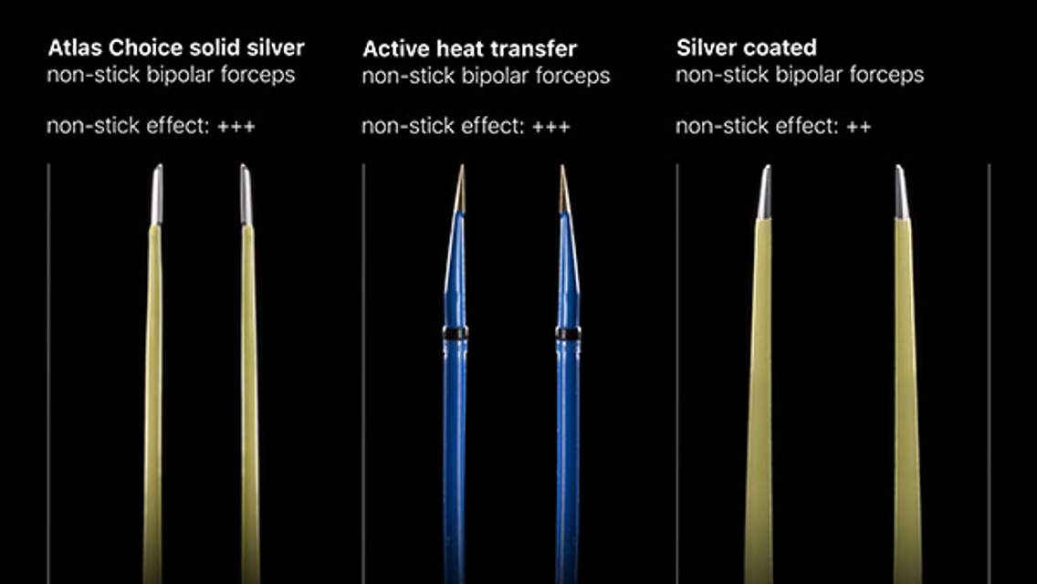
- Next, using bipolar electrocautery and microscissors, I dissect, coagulate, and divide the fine feeding vessels clearly leading to the capsule.
- Blunt dissection via a fine dissector allows mobilization of the capsule away from the gliotic margins while maintaining the capsule integrity.
- The CM can be removed en bloc or piecemeal (using pituitary rongeurs), depending on the available exposure trajectory. Most spinal CMs demand piecemeal removal via a small incision on the surface of the cord.
- Gentle coagulation of the resection walls may be necessary for complete hemostasis. Tamponade using small pieces of thrombin-soaked cotton may avoid any coagulation injury.
- Careful inspection of the resection cavity is mandatory. The color of portions of the CM may appear similar to that of the gliotic margins. Fine forceps may be used to draw upon suspicious material. Developmental venous anomalies (DVAs) are left intact.
- Gliotic borders are left intact to avoid neurologic consequences.
These important steps are further illustrated below.
Figure 2: After exposure of the CM, a suction device or pituitary rongeurs are used to evacuate the hematoma from within and around the CM (A). Next, the small feeding vessels are isolated, coagulated, and cut (B). The capsule of the lesion is then mobilized away from the gliotic margins (C). The DVA is carefully protected and mobilized away from the lesion (D). Finally, the CM is removed.
Postoperative Considerations
Some patients may suffer transient neurologic worsening. Patients should undergo MR imaging postoperatively to confirm gross total resection of their mass and establish a new imaging baseline for future comparison. Further follow-up imaging is up to the discretion of the surgeon. I tend to repeat MR imaging in 3-month postoperatively and every 3-5 years thereafter unless new neurologic deficits develop.
Pearls and Pitfalls
- Spinal cord CMs are frequently recognized as a cause of chronic myelopathy or acute focal neurologic deficits.
- Symptomatic patients should undergo microsurgery with a goal of gross total resection.
- Associated hemosiderin-stained tissues and venous developmental anomalies should be preserved.
References
Badhiwala JH, Farrokhyar F, Alhazzani W, Yarascavitch B, et al. Surgical outcomes and natural history of intramedullary spinal cord cavernous malformations: A single-center series and meta-analysis of individual patient data. J Neurosurg Spine. 2014; 21:662-676.
Jallo GI, Freed D, Zareck M, Epstein F, Kothbauer KF. Clinical presentation and optimal management for intramedullary spinal cavernous malformations. Neurosurg Focus. 2006; 21:e10.
Kalani MY, Kalani MA, Spetzler RF. Microsurgery of intramedullary spinal cavernous malformations, in Spetzler RF, Kalani MY, and Nakaji P, (eds): Neurovascular Surgery. Second edition. New York: Thieme Medical Publishers; 2015:448-454.
Kshettry VR, Healy AT, Jones NG, Mroz TE, Benzel EC. A quantitative analysis of approaches to the ventral thoracic spinal canal. Spine J. 2015; Epub ahead of print.
Please login to post a comment.


