Progressive Multifocal Leukoencephalopathy
Progressive Multifocal Leukoencephalopathy (PML) is the infection of myelin-producing oligodendrocytes by reactivated JC virus due to immunodeficiency, particularly in AIDS patients when the CD4+ cell count falls below 100 cells/µl. 90% of patients die of the disease within 1 year of diagnosis.
Imaging Features
PML shares similarities to other demyelinating diseases – including the presence of a leading edge enhancement and restricted diffusion, its predominantly white matter distribution, and central edema. The distribution of PML is classically asymmetric (while HIV encephalitis is classically symmetric). Though PML may involve most areas of the brain, common locations include subcortical white matter (often frontoparietal) and thalami, progressing to involve more and more areas of the brain in a confluent and often multifocal pattern.
- CT:
- Asymmetric focal low-density lesions in the periventricular and subcortical white matter.
- MRI:
- T1: Low signal intensity, often quite striking.
- T2/FLAIR: High signal focal white matter lesions.
- SWI/GRE: Usually normal. Minimal hemorrhage is sometimes seen and presents as patchy low signal.
- T1+C: Usually non-enhancing. If present, it appears as mild peripheral enhancement. Interestingly, the presence of enhancement is a good prognostic factor – one study found that 50% of long-term survivors had enhancing lesions, while only 9% of short-term survivors did.
- DWI/ADC: Restricted diffusion in the periphery (high DWI signal, low ADC signal) is suggestive of a “leading edge” of demyelination. The center of the lesions usually shows T2 shine-through reflecting edema (bright DWI and ADC signal).
- Spectroscopy: Decreased NAA. Increased lactate and lipids, and mildly elevated choline.
Figure 1: PML on Contrast-enhanced CT: This 43-year-old male with AIDS and CD4 <13 demonstrates left parietal juxtacortical hypodensity without enhancement (left). Right greater than left corpus callosum splenium hypodensity also indicates involvement in this area.
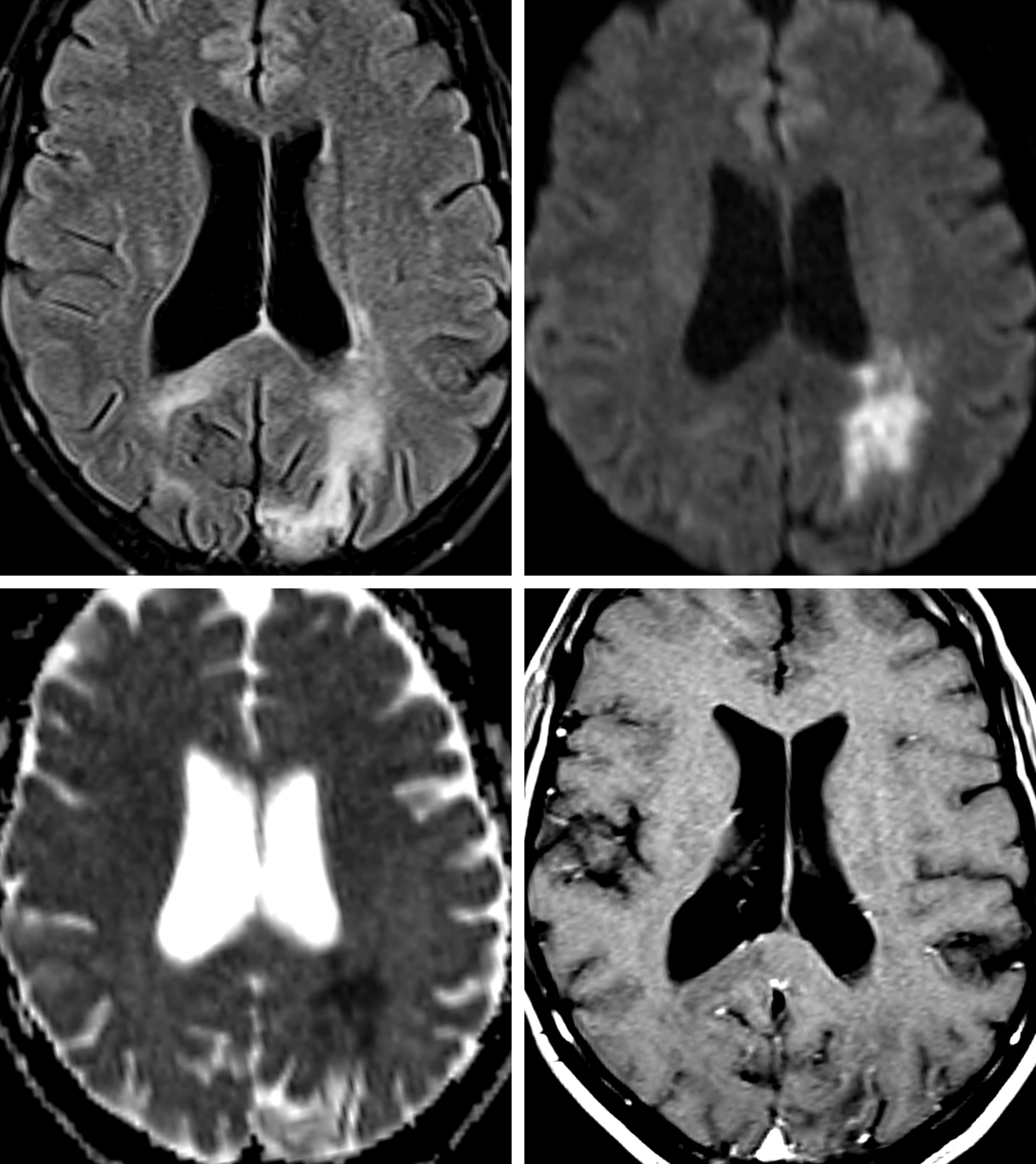
Figure 2: In the same patient as in Figure 1, the FLAIR sequence (top row left) shows both hyperintense and hypointense (due to CSF-like signal suppression) signal intensities in the areas of PML involvement. DWI (top row right) and ADC (bottom row left) images show areas of restricted diffusion in areas of PML involvement, a finding that is sometimes present. T1+C (bottom row right) illustrates striking T1 hypointensity in the subcortical white matter involvement of the posterior parietal lobe, with only minimal adjacent enhancement.
Figure 3: 3-year-followup MRI for this same patient after successful treatment for HIV illustrates the long-term damage done by PML. The encephalomalacia and gliosis in the left parietal lobe demonstrates areas of parenchymal hyperintensity on FLAIR with CSF-like hypointensity in the areas of most severe chronic involvement more posteriorly. T1-weighted image (right) also illustrates the atrophy and striking hypointensity.
Differential Diagnosis
- AIDS dementia complex (AKA HIV encephalopathy)
- Diffuse, symmetric bilateral white matter increased FLAIR signal and atrophy with sparing of the subcortical U-fibers (white matter immediately adjacent to cortex) until late in the disease
- Demyelinating disorders of other causes
- Imaging appearance, patient demographics, clinical scenario and acuity of the disease can be helpful in differentiating PML from MS or acute disseminated encephalomyelitis (ADEM)
- MS usually shows a greater number of smaller lesions than PML
- ADEM appearance can have significant overlap, but usually has a more “flocculent”, cloud-like appearance than PML
- Imaging appearance, patient demographics, clinical scenario and acuity of the disease can be helpful in differentiating PML from MS or acute disseminated encephalomyelitis (ADEM)
For more information, please see the corresponding chapter in Radiopaedia and the PML tumor mimic chapter in the Neurosurgical Atlas.
Contributor: Jordan McDonald, MD
References
Smith AB, Smirniotopoulos JG, Rushing EJ. From the archives of the AFIP – Central nervous system infections with human immunodeficiency virus infection: Radiologic-pathologic correlation. Radiographics. 2008; 28:2033-2058.
Berger JR, Levy RM, Flomenholt D, Dobbs M. Predictive factors for prolonged survival in acquired immunodeficiency syndrome-associated progressive multifocal leukoencephalopathy. Ann Neurol. 1998; 44(3): 341-349.
Whiteman ML, Post MJ, Berger JR, et al. Progressive multifocal leukoencephalopathy in 47 HIV-seropositive patients: Neuroimaging with clinical and pathologic correlation. Radiology. 1993; 187(1): 233-240.
Please login to post a comment.


