Aneurysms of the Posterior Inferior Cerebellar Artery
Abstract
The clinical and anatomical features of 21 surgically treated saccular aneurysms of the posterior inferior cerebellar artery (PICA) are analyzed. Seventeen of these lesions originated from the PICA-vertebral junction, and four arose from distal PICA branching sites. Twelve lesions arose from the left PICA, nine were right-sided, and all were small (less than 12.5 mm). Most of these aneurysms occurred in females (16 of 21) and presented as classic subarachnoid hemorrhage. The lack of specific focal deficits prevented an accurate pre-angiographic determination of aneurysm location in most instances. Clinically significant vasospasm and aneurysm multiplicity occurred with approximately equal frequency as at other locations.
The angiographic and surgical features of these lesions are determined by the course of the vertebral artery and PICA; that is, they occur at branching sites and at curves in the parent vessel, and point in the direction in which flow would have continued if the curve at the aneurysm's origin had not been present. Aneurysms at the PICA-vertebral junction usually occur at least 1 cm above the foramen magnum level, arise distal to the PICA origin in the angle between the two vessels, and are best approached by a paramedian incision with the patient in the lateral recumbent position. Isolated clipping of the aneurysm neck is essential in this instance, as trapping may compromise vital perforating arteries of the brain stem. More distal (retromedullary) PICA aneurysms are sometimes associated with another vascular anomaly (two cases in this series), and are best handled through a bilateral suboccipital craniectomy. Clipping of the neck is the preferred treatment, but trapping is usually safe, if necessary.
Introduction
Vertibrobasilar aneurysms constitute approximately 15% of all intracranial aneurysms, most of which arise from the basilar apex.21 One-fifth of these posterior fossa lesions originate from the posterior inferior cerebellar artery (PICA), thus accounting for 3% of all intracranial aneurysms. The anatomy of PICA is quite variable,16 and aneurysms from this artery are often tightly confined between the medulla oblongata, the anterolateral base of the skull, and the lower cranial nerves. A clinical and anatomical correlation of our experience with 21 surgically treated PICA aneurysms forms the basis for this report.
Summary of Cases
Clinical Data
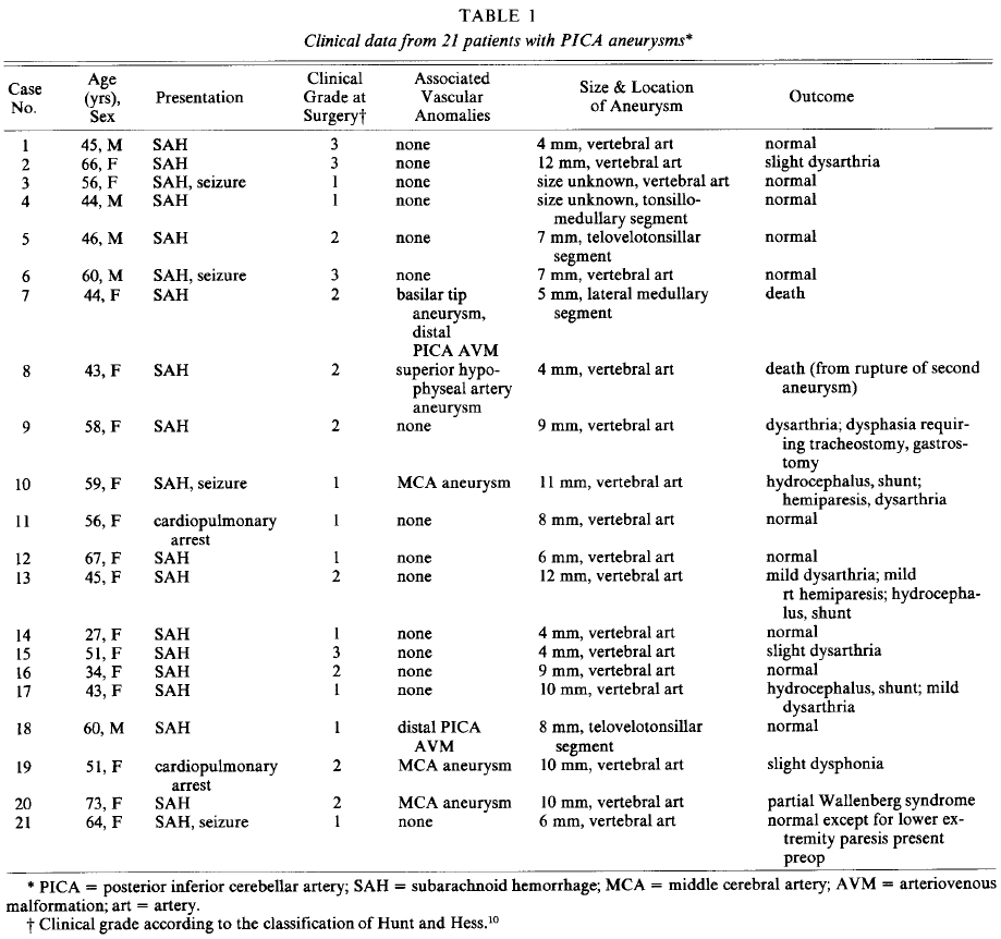
Between 1972 and 1982, 21 cases of saccular PICA-vertebral and distal PICA aneurysms were operated on at the University of Florida (Table 1). The average age of these patients was 52 years, with a range of 27 to 73 years, and there was a striking female predominance (16 patients). All 21 patients were initially evaluated at other hospitals, and subsequently transferred to our institution. The mean time from ictus to referral was 8.2 days, with a range of several hours to 22 days, excluding two patients. One of these was initially operated on unsuccessfully elsewhere and was admitted 62 days after ictus, and the other was seen 1 year after subarachnoid hemorrhage (SAH). This delay in referral skews our series toward patients who did relatively well after their initial hemorrhage, and prevents any conclusions regarding the natural history and preoperative mortality rate associated with such lesions.
All patients presented with classic SAH symptoms: sudden severe headache (usually occipital) followed by an altered level of consciousness (19 cases), including lethargy or coma. Concomitant with their ictus, four patients had seizures and two had a cardiopulmonary arrest. Twelve patients exhibited no focal abnormalities during the initial evaluation. The remaining patients exhibited some focal neurological deficits, including lower extremity dysesthesia in two cases, paresis in one lower extremity in one case, oculomotor dysfunction in three cases (Horner's syndrome in one, abducens palsy in one, and entropion, poorly classified, in one), bilateral Babinski signs in one case, and dysarthria in three cases. One dysarthric patient also had a hemiparesis, which cleared prior to admission. A history of hypertension was present in eight patients. Lumbar puncture revealed bloody or xanthochromic cerebrospinal fluid (CSF) in all cases.
Radiographic Data
Computerized Tomography
Of the eight patients who underwent computerized tomography (CT) scanning within 24 hours of their ictus, six had evidence of blood in the subarachnoid space or ventricles (Table 1). In two cases, the hemorrhage was noted only in the basilar cisterns. The other four cases, however, demonstrated blood within the ventricular system, in one affecting the fourth ventricle only, in one confined to the third and fourth ventricles (with basilar subarachnoid clot), and in two throughout all the ventricles. The patient with an isolated fourth ventricle hematoma also had acute hydrocephalus. Three patients in our series ultimately required ventriculoperitoneal shunting for persistent and symptomatic hydrocephalus.
FIG. 1. Normal anatomy of the posterior inferior cerebellar artery (PICA). Left: Lateral view of the brain stem and cerebellum demonstrating the normal course of the PICA and its division into five segments (A-E). The anterior medullary segment (A) begins at the PICA-vertebral junction and becomes continuous with the lateral medullary segment (B) as it courses rostrally, caudally, or through the rootlets of the hypoglossal nerve. The lateral medullary segment extends to the rootlets of the glossopharyngeal, vagal, and accessory nerves. The tonsillomedullary segment (C) extends from these rootlets and often forms a caudally convex loop (caudal loop). It then turns rostrally and at the midportion of the tonsil becomes continuous with the telovelotonsillar segment (D). This segment frequently has a rostrally convex loop (cranial loop) near the roof of the fourth ventricle. The telovelotonsillar segment then runs through a cleft between the vermis medially and the tonsil and cerebellar hemispheres laterally to become continuous with the cortical segment (E). This segment divides into a medial branch which passes posteriorly and superiorly close to the midline to supply the vermis, and lateral branches, which pass laterally to supply the inferior surface of the cerebellar hemispheres. Right: Posterior view. Note the intimate relationship of the lateral medullary segment (B) to rootlets of the ninth, 10th, and 11 th cranial nerves. The anterior medullary (A), lateral medullary, and tonsillomedullary (C) segments give off brain-stem perforators. The telovelotonsillar segment is in close proximity to the roof of the fourth ventricle. Cortical segments (E) begin as the PICA passes out of the fissure between the vermis medially and the tonsil and cerebellar hemispheres laterally.
FIG. 2. Distribution of the 21 aneurysms along the course of the posterior inferior cerebellar artery (PICA). Seventeen were at the PICA-vertebral artery junction, one on the lateral medullary segment, one on the tonsillomedullary segment (caudal loop), and two on the telovelotonsillar segment (cranial loop).
Angiography
All patients underwent at least threevessel cerebral angiography (Table 1). If there was not adequate reflux of contrast material to fill the contralateral vertebral artery to the origin of the PICA, the fourth vessel was also injected. The normal anatomy of the PICA is outlined in Fig. 1, and the distribution of our series of aneurysms along its course is outlined in Fig. 2. Seventeen patients had aneurysms located at the PICA-vertebral junction, with the aneurysm arising just beyond the PICA origin in the angle between the two vessels. Four aneurysms were located more distally along the PICA, with one arising from the lateral medullary segment, one from the retromedullary segment (caudal loop), and two from the telovelotonsillar segment (cranial loop). The aneurysms ranged in size between 2 and 12 mm, with an average size of 7 mm (the size of two was unknown). Twelve arose from the left, and nine from the right PICA circulations. No statistically significant correlation between laterality and hypertension could be identified (four of eight hypertensive patients had left-sided aneurysms).
Associated vascular anomalies were noted in six cases (Table 1). One patient had an aneurysm of the contralateral superior hypophyseal artery; three had middle cerebral artery (MCA) aneurysms, two contralateral, and the other ipsilateral to the PICA aneurysm. In each of these four cases, the PICA aneurysm was located at the PICA-vertebral junction. The remaining two patients had ipsilateral arteriovenous malformations (AVM's) supplied primarily by the PICA containing the aneurysm. In each of these cases, the aneurysm was located more distally along the PICA course. One of these patients with AVM's also had a 16-mm aneurysm at the basilar apex.
Angiographic evidence of vasospasm was found in seven cases. Two patients had local spasm only (on the ipsilateral vertebral artery), and both were mildly disoriented. Two had local spasm of the vertebral artery and also distal spasm, one severe enough to delay surgery. Three patients had distal spasm only, two of them postoperatively. They were both left with minor residual neurological deficits, probably secondary to ischemia.
One patient (Case 13) underwent attempted wrapping of her aneurysm at another hospital 2 months before her admission to our institution. Repeat angiography showed an increase in the size of the saccular PICA-vertebral junction aneurysm and a questionable associated dissection of the vertebral artery.
The necessity of visualizing all four vessels at angiography 7,24 was emphasized in two of our cases. One patient (Case 3) had a reportedly normal angiogram following SAH, but the initial study had not included visualization of the PICA-vertebral junction from which her aneurysm arose. The other patient (Case 10) was diagnosed as having a ruptured MCA aneurysm after bilateral carotid angiography, but subsequent four-vessel angiography and surgery demonstrated that the aneurysm that had ruptured originated from her left PICA.
Treatment
Preoperative Care
All patients were begun on Amicar (aminocaproic acid, 36 to 48 gm/day) upon arrival at out institution, except for one patient admitted 1 year after her ictus. Other standard procedures consisted of bed rest in a quiet room, blood pressure control if necessary, and occasional use of phenobarbital for sedation. In more recent cases, kanamycin and reserpine were also added if the patients were received within 1 week of their hemorrhage.
No patient had recurrent hemorrhage after arrival at our institution, but two had episodes before transfer. One was a patient who had bled 7 years prior to admission and was found after her second bleed to have a PICA aneurysm. Another patient had a second ictal event 7 days after her initial hemorrhage which was presumed to be recurrent bleeding. Lumbar puncture was not repeated until 2 days later, however, revealing only xanthochromic CSF.
Surgery
Average time from the most recent SAH to surgery was 11 days, excluding one interval of 90 days (Case 13) and another of 1 year (Case 21). Suboccipital craniectomy was performed in 20 cases, and a subtemporal craniectomy in one. Eight procedures were conducted with the patient sitting, 11 in the lateral recumbent "park bench" position, one prone, and one in the straight lateral position. Spinal drainage, mannitol, and hyperventilation were used to decrease retraction pressure in each case. The operating microscope was used in 20 cases, and surgical loupes in the remaining case. All aneurysms were clipped across their necks, except for the one with the possible dissecting component, in which the vertebral artery was clipped proximal to the PICA.
Results
Results were divided into four categories: 1) good, able to return to full previous activities; 2) fair, minor neurological deficits which slightly modified life-style (such as functionally significant dysarthria, decreased palatal excursion); 3) poor, disabling neurological deficits; and 4) death. Thirteen patients had good results, including 10 who were neurologically normal at follow-up examination, and three with an insignificant dysphonia or dysarthria. Four patients had fair results; in three of these, deficits were due to cranial nerve palsy causing functionally significant dysarthria. These deficits were presumed due to nerve damage during surgical manipulation, although the effect of vasospasm and small PICA infarctions cannot be eliminated. The other fair result was in a patient who had temporary clipping of both the vertebral artery and PICA when her aneurysm tore off across the neck during attempted clipping. Postoperatively, she developed a partial Wallenberg syndrome (decreased gag reflex, hoarseness, upper extremity dysmetria, and alternating decreased pain sensation) secondary to PICA ischemia. These deficits are continuing to improve at this time.
Two patients had poor results. One of these patients was ambulatory, but exhibited multiple cranial nerve dysfunction from a large PICA infarction. Her palatal dysfunction led to recurrent aspiration pneumonia requiring a tracheostomy and gastrostomy. Another patient had persistent mild hemiparesis and multiple cranial nerve dysfunction also from ischemic damage to the PICA territory. In both instances, these deficits appeared shortly after surgery, and their cause was unclear.
There were two deaths in this series. One of these patients (Case 7) had a basilar tip aneurysm and cerebellar AVM as well as a PICA aneurysm. First, the basilar artery aneurysm was approached via a subtemporal craniotomy. Several weeks later, however, she died of a postoperative hematoma following a suboccipital craniectomy for clipping of the PICA aneurysm and removal of the AVM. The other patient who died (Case 8) had both a PICA aneurysm and a superior hypophyseal artery aneurysm. She underwent uneventful clipping of her PICA aneurysm, but died 2 years later from SAH secondary to rupture of the supratentorial aneurysm. Angiography prior to her death revealed excellent obliteration of the PICA aneurysm.
Discussion
Historical Perspective
Although Cruveilhier described a spherical aneurysm arising from the PICA-vertebral junction in 1829,4 it was not until 1947 that Rizzoli and Hayes23 performed the first surgical procedure on an aneurysm known to arise from this vessel. In their case, preoperative angiography was not done, and the posterior fossa location of the lesion was deduced from a shift of the fourth ventricle seen at ventriculography. The aneurysm was trapped between silver clips.
Richardson's study22 of the natural history of aneurysms following SAH revealed that those arising from the vertebrobasilar system were associated with the highest mortality rate. Uihlein and Hughes26 reported a series of 14 posterior fossa aneurysms treated without definitive surgery; eight of these patients died of aneurysm rupture. Routine utilization of vertebral angiography after normal carotid angiography in patients with SAH enhanced the recognition of these aneurysms. In 1958, DeSaussure, et al.,5 reported the successful trapping of two PICA aneurysms that had been defined by preoperative angiography. Further refinements, including transfemoral catheterization, subtraction, and magnification, have enhanced the preoperative angiographic assessment of size, location, and position of such aneurysms.
In 1967, Rand and Jannetta19 recognized the benefit of the operating microscope for aneurysms of the vertebrobasilar system, both for the delicate dissection of the neck of the aneurysm and for preventing inadvertent occlusion of the small perforating arteries at the time of clipping. The excellent results enjoyed by Drake7 and others confirm these benefits.
Clinical Characteristics
This series of PICA-vertebral and distal aneurysms represents the experience of four neurosurgeons with these lesions over a 10-year period. In this group of 21 patients, there was approximately a 3:1 preponderance of females. Several other series of PICA aneurysms demonstrate a similar female predisposition,3,11,24 while one shows an equal male to female ratio.6 Only the series of Hammon and Kempe shows a male predominance, a skew not unexpected from their predominantly military population at Walter Reed General Hospital.
The average age in our series (52 years) agreed with that found by others,3,11,24 and was essentially the same as that found in the Cooperative Study17 for all aneurysmal SAH. The Hammon and Kempe series8 differs in that the average age of patients with PICA aneurysms is 33 years, and again probably reflects their specialized referral base.
Although Laine15 described a syndrome of localizing value for ruptured vertebral and PICA aneurysms, this was not confirmed in our series. All patients had syncopal episodes or seizures, but none had a classic "drop attack." Two patients had dysesthesiae, three had dysarthria, and one had a sixth nerve paresis. The most common presentation in our series, as well as in others,7,24,25 was headaches, decreased level of consciousness, and meningismus without focal deficits.
All aneurysms in our patients were less than 1.2 cm in size. This is consistent with Drake's observation7 that most of these aneurysms are less than 1.25 cm or greater than 2.5 cm. We did not, however, see the large percentage of giant aneurysms reported by Drake7 (six of 50 aneurysms) and Kempe14 (15 of 48). Even when these aneurysms reach giant proportions, the clinical characteristics are quite variable, and these lesions have been reported to present as posterior fossa tumor, 12 foramen magnum syndrome, 13 obstructive hydrocephalus, 1 and cerebellopontine angle syndrome.2
Vasospasm (combined angiographic and clinical) was found in seven patients in this study. This incidence of 33% is essentially the same as that reported for all aneurysms.18
Multiple aneurysms were found in five of our 21 patients (24%), correlating well with the number found in the literature for aneurysms as a whole.21 The rare association of a PICA aneurysm and AVM fed primarily by the PICA was also found in two patients. As in the case reported by Higashi, et al.,9 the aneurysms in our cases were located along the distal course of the PICA rather than at the PICA-vertebral junction.
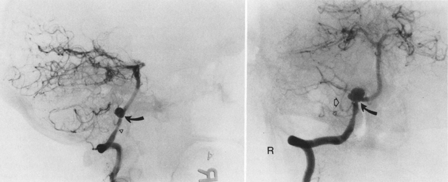
FIG. 3. Left: Subtracted right vertebral angiogram, lateral projection, demonstrating an aneurysm (curved arrow) arising from the junction of the right posterior inferior cerebellar artery (PICA) and the right vertebral artery. In this projection, this 11-mm aneurysm projects cephalad and upward toward the anterolateral surface of the medulla. There is mild arterial spasm (arrowhead) of the intradural segment of the right vertebral artery immediately preceding the PICA origin. Right: Subtracted right vertebral angiogram, anteroposterior transfacial projection, illustrating the orientation of the PICA-vertebral aneurysms relative to the medially directed vertebral artery and the laterally oriented PICA. The aneurysm (curved arrow) projects cephalad. Additional arterial spasm is present in the lateral medullary segment of the PICA (open arrow).
Surgical Principles
The PICA has the most variable course of all the cerebellar arteries, and its course (recently reviewed by Lister, et al.16) was the primary determinant of the location of the aneurysm and of the direction in which the aneurysm was pointed. These lesions followed the habit of aneurysms, set forth by Rhoton, 20 in that they occurred at branching points and at curves, and pointed in the direction that the blood flow would have taken if the curve had not been present.
PICA-Vertebral Aneurysms
Seventeen of our 21 aneurysms were located at the PICA-vertebral junction (Fig. 2). A compilation of data from several other large series of PICA aneurysms suggests that approximately two-thirds of all such lesions will arise at the PICA-vertebral junction, while one-third will occur more distally.
The PICA-vertebral junction aneurysms usually arose at a curve where the vertebral artery turned medially to join the contralateral vertebral artery. Thus, they tended to arise just above the PICA origin in the angle between the vertebral artery and PICA, usually pointing superiorly and often partially embedded into the anterolateral medulla (Fig. 3). In the normal course of the PICA,16 this point usually lies 10 mm above the foramen magnum in the anterolateral subarachnoid space between the medulla, skull base, and lower cranial nerves.
The point at which the aneurysm originates determines the surgical approach and alternatives in aneurysm obliteration. Aneurysms at the PICA-vertebral junction and the initial two segments of the PICA would best be approached via a paramedian incision to afford the best visualization of their necks. Trapping procedures should not be used on these aneurysms, as blood flow to vital medullary perforators may be compromised.
The early surgical procedures in this series of patients were done by several neurosurgeons, usually with the patient in a sitting position. In our more recent operations (that is, in the last 11 cases), the patient was placed in the lateral recumbent "park bench" position. This approach appears advantageous in PICA-vertebral junction aneurysms for several reasons: 1) there is a decreased incidence of air embolism and hypotension, because the head is lower than the heart; 2) less retraction on the cerebellum (and if necessary, the medulla) is necessary, as gravity causes them to fall away; and 3) the potential obstructive effect of the jugular tubercle and foramen magnum can be minimized by the superior and more lateral angle of this exposure.
A major cause of morbidity in PICA-vertebral aneurysms is the inadvertent disruption of the rootlets of the ninth through the 12th cranial nerves. The PICA takes a variable course through these rootlets, frequently looping superiorly so that it touches the anterior or inferior surface of the facial and/or vestibulocochlear nerve.16 With the patient in the "park bench" position, the rostral location of the surgeon allows better visualization of proximal aneurysms in this area, and minimizes the amount of retraction and dissection necessary for excellent visualization and clip placement.
The extreme tortuosity of the vertebral and PICA arteries may also occasionally influence the laterality of the operative approach. Drake7 described an aneurysm arising from the right vertebral artery distal to the PICA which was clipped through a left suboccipital craniectomy because the tortuosity of the vertebral artery caused it to cross the midline.
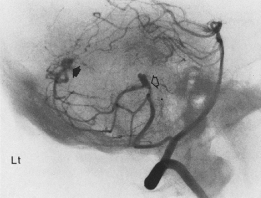
FIG. 4. Subtracted left vertebral angiogram, lateral projection, demonstrating the presence of an 8-mm aneurysm (open arrow) arising at the junction between the retromedullary and telovelotonsillar segment of the left posterior inferior cerebellar artery. This corresponds topographically to the region of the contiguous superior tonsillar pole and posterior lateral recess of the fourth ventricle. In addition, a small arteriovenous malformation (solid arrow) is present along the superior surface of the vermis.
Distal PICA Aneurysms
Four of our aneurysms arose from distal segments of the PICA, including one from the lateral medullary segment, one from the tonsillomedullary segment, and two from the telovelotonsillar segment (Fig. 2). As with other aneurysms, these lesions arise at branching sites at a curve of the parent vessel, and point in the direction that the vessel would have pointed if the curve had not been present (Fig. 4). Two of these cases had ipsilateral AVM's fed primarily by the PICA containing the aneurysm, a high incidence which strongly supports a "flowrelated" origin of aneurysms arising along the distal course of cerebral vessels.
Aneurysms from the first two segments of the PICA are best approached via a lateral exposure, but those arising from the distal three segments, posterior to the brain stem, are better handled through a bilateral suboccipital craniectomy. Although clipping across the aneurysmal neck is preferable, trapping may be utilized in those lesions arising from or distal to the telovelotonsillar segment, as no further brain-stem perforators arise beyond this point.
This article was originally published here: Hudgins RJ, Day AL, Quisling RG, Rhoton AL Jr., Sypert GW, Garcia-Bengochea F. Aneurysms of the posterior inferior cerebellar artery: a clinical and anatomical analysis. J Neurosurg 1983;58:381–387, doi.org/10.3171/jns.1983.58.3.0381, and is included through an exclusive partnership with the Journal of Neurosurgery and its parent company, the American Association of Neurological Surgeons (AANS). The AANS retains full copyright. The appearance of this material here does not imply open access or free use by any other party.
The Neurosurgical Atlas is honored to maintain the legacy of Albert L. Rhoton, Jr, MD
References
- Alexander E Jr, Davis CH Jr, Pikula L: Aneurysm of the posterior inferior cerebellar artery filling the fourth ventricle. J Neurosurg 24:99-101, 1966
- Bull J: Massive aneurysms at the base of the brain. Brain 92:535-579, 1969
- Chou SN, Ortiz-Suarez H J: Surgical treatment of arterial aneurysms of the vertebrobasilar circulation. J Neurosurg 41:671-680, 1974
- Cruveilhier J: Anatomie Pathologique de Corps Humain. Paris: JB Bailliere, 1829-1835, Vol 2. Cited in Schwartz HG: Arterial aneurysm of the posterior fossa. J Neurosurg 5:312-316, 1948
- DeSaussure RL, Hunter SE, Robertson JT: Saccular aneurysms of the posterior fossa. J Neurosurg 15: 385-391, 1958
- Dimsdale H, Logue V: Ruptured posterior fossa aneurysms and their surgical treatment. J Neurol Neurosurg Psychiatry 22:202-217, 1959
- Drake CG: Treatment of aneurysms of the posterior cranial fossa. Prog Neurol Surg 9:122-194, 1978
- Hammon WM, Kempe LG: The posterior fossa approach to aneurysms of the vertebral and basilar arteries. J Neurosurg 37:339-347, 1972
- Higashi K, Hatano M, Yamashita T, et al: Coexistence of posterior inferior cerebellar artery aneurysm and arteriovenous malformation fed by the same artery. Surg Neurol 12:405-408, 1979
- Hunt WE, Hess RM: Surgical risk as related to time of intervention in the repair of intracranial aneurysm. J Neurosurg 28:14-20, 1968
- Jamieson KG: Aneurysms of the vertebro-basilar system. Surgical intervention in 19 cases. J Neurosurg 21:781-797, 1964
- Jane JA: A large aneurysm of the posterior inferior cerebellar artery in a l-year-old child. J Neurosurg 18:245-247, 1961
- Judice D, Connolly ES: Foramen magnum syndrome caused by a giant aneurysm of the posterior inferior cerebral artery. Case report. J Neurosurg 48:639-641, 1978
- Kempe LG: Aneurysms of the vertebral artery, in Pia HW, Langmaid C, Zierski J (eds): Cerebral Aneurysms. Advances in Diagnosis and Therapy. Berlin/Heidelberg/New York: Springer-Verlag, 1979, pp 119-120
- Laine E: Arterial vertebro-basilar aneurysms. Prog Brain Res 30:323-346, 1968
- Lister JR, Rhoton AL Jr, Matsushima T, et al: Microsurgical anatomy of the posterior inferior cerebellar artery. Neurosurgery 10:170-199, 1982
- Locksley HB: Report on the Cooperative Study of Intracranial Aneurysms and Subarachnoid Hemorrhage. Section V, Part I. Natural history of subarachnoid hemorrhage, intracranial aneurysms, and arteriovenous malformations. Based on 6368 cases in the Cooperative Study. J Neurosurg 25:219-239, 1966
- Mohan J: The neurosurgeon's view, in Boullin DJ (ed): Cerebral Vasospasm. New York: John Wiley and Sons, 1980, pp 15-35
- Rand RW, Jannetta PJ: Micro-neurosurgery for aneurysms of the vertebral-basilar artery system. J Neurosurg 27:330-335, 1967
- Rhoton AL Jr" Anatomy of saccular aneurysms. Surg Neurol 14:59-66, 1980
- Rhoton AL Jr, Jackson FE, Gleave J, et al: Congenital and traumatic intracranial aneurysms. CIBA Clin Syrup 29(4):2-40, 1977
- Richardson AE: The natural history of patients with intracranial aneurysms after rupture. Prog Brain Res 30:269-273, 1968
- Rizzoli HV, Hayes G J: Congenital berry aneurysm of the posterior fossa. Case report with successful operative excision. J Neurosurg 10:550-55 l, 1953
- Rothman SLG, Azarkia B, Kier EL, et al: The angiography of posterior inferior cerebellar artery aneurysms. Neuroradiology 6:1-7, 1973
- Sharr MM, Kelvin FM: Vertebrobasilar aneurysms. Experience with 27 cases. Eur Neurol 10:129-143, 1973
- Uihlein A, Hughes RA: The surgical treatment ofintracranial vestigial aneurysms. Surg Clin North Am 35:1071-1083, 1955
Please login to post a comment.


