Central Neurocytoma
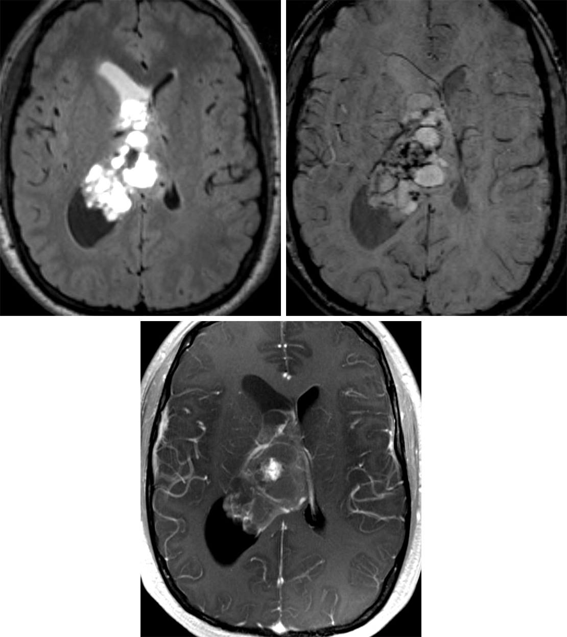
Figure 1: (Top Left) This large central neurocytoma demonstrates its typically cystic feature on axial FLAIR. This image also shows an entrapped right frontal horn indicated by incomplete suppression of cerebrospinal fluid. (Top Right) The SWI image shows dark susceptibility artifact that likely represents calcifications commonly seen in these tumors. (Bottom) Central neurocytomas typically have some enhancement but less than many other tumors, as seen on this postcontrast T1WI.
Figure 2: (Top Left) Axial T1-weighted postcontrast image demonstrates a heterogeneously enhancing mass in the left lateral ventricle, which appears inseparable from the septum pellucidum. (Top Right) T2-FS image demonstrates the cystic and bubbly appearance of this mass. Enhancing components correspond to regions of restricted diffusion and presumed hypercellularity in diffusion-weighted imaging (bottom left) and ADC (bottom right). The pathology is consistent with central neurocytoma.
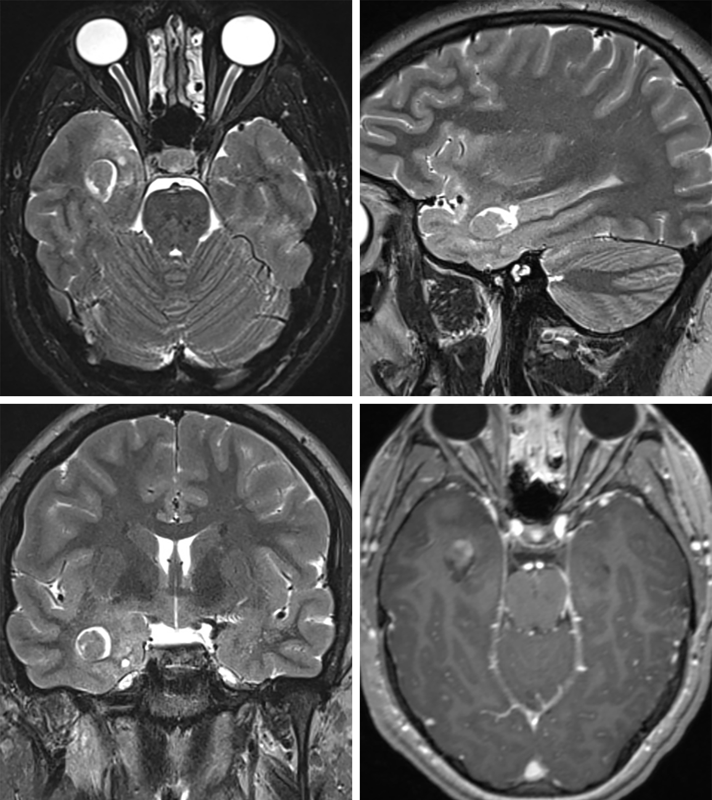
Figure 3: This right temporal extraventricular neurocytoma mimics the appearance of other low-grade primary brain tumors such as ganglioglioma and DNET. It demonstrates T2 hyperintense (top left, axial; top right, sagittal; bottom left, coronal) nonenhancing (bottom right) cystic components and intermediate signal intensity (top left, top right, and bottom left) mildly enhancing (bottom right) solid component, similar to the more common intraventricular central neurocytoma.
BASIC DESCRIPTION
- Well-marginated, usually benign intraventricular neuroepithelial tumor
PATHOLOGY
- WHO grade II
- Higher-grade atypical, anaplastic variant is rare
- Round cells with stippled nuclei are characteristic microscopic features
- Electron microscopy: synaptophysin positive
CLINICAL FEATURES
- Affects all ages (commonly third decade of life)
- No gender predilection
- Commonly presents with signs/symptoms of increased intracranial pressure secondary to obstructive hydrocephalus
- Headache, nausea, vomiting, and altered mental status
- Treatment: surgical resection; chemoradiation or radiosurgery for unresectable tumors
- Prognosis: recurrence uncommon after total resection; 5-year survival rate, 90%
IMAGING FEATURES
- General
- Supratentorial intraventricular mass with cystic or bubbly appearance
- Often located with lateral ventral frontal horn or body near the foramen of Monro
- Attachment to the septum pellucidum or lateral ventricular wall
- ±Third ventricular extension
- Frequently shows calcification
- CT
- Heterogeneous density intraventricular mass due to solid and cystic components
- ±Calcification
- Heterogeneous enhancement on contrast-enhanced CT
- MRI
- T1WI: heterogeneous but predominantly isointense
- T2WI: usually hyperintense with cystic or bubbly appearance; ±flow voids
- FLAIR: heterogeneously hyperintense
- T2*/GRE/SWI: black signal blooming in foci of calcification
- T1WI+C: heterogeneous enhancement
- MRS: increased Cho, decreased NAA; glycine peak at 3.55
IMAGING RECOMMENDATIONS
- MRI with contrast, including coronal T2WI, to better detect tumor attachment site
For more information, please see the corresponding chapter in Radiopaedia.
Contributors: Rachel Seltman, MD, and Jacob A. Eitel, MD
References
Donoho D, Zada G. Imaging of central neurocytomas. Neurosurg Clin N Am 2015;26:11–19. doi.org/10.1016/j.nec.2014.09.012.
Kocaoglu M, Ors F, Bulakbasi N, et al. Central neurocytoma: proton MR spectroscopy and diffusion weighted MR imaging findings. Magn Reson Imaging 2009;27:434–440. doi.org/10.1016/j.mri.2008.07.012.
Osborn AG, Salzman KL, Jhaveri MD. Diagnostic Imaging (3rd ed). Elsevier, Philadelphia, PA; 2016.
Patel DM, Schmidt RF, Liu JK. Update on the diagnosis, pathogenesis, and treatment strategies for central neurocytoma. J Clin Neurosci 2013;20:1193–1199. doi.org/10.1016/j.jocn.2013.01.001.
Schmidt MH, Gottfried ON, von Koch CS, et al. Central neurocytoma: a review. J Neurooncol 2004;66:377–384. doi.org/10.1023/b:neon.0000014541.87329.3b.
Tlili-Graiess K, Mama N, Arifa N, et al. Diffusion weighted MR imaging and proton MR spectroscopy findings of central neurocytoma with pathological correlation. J Neuroradiol 2014;41:243–250. doi.org/10.1016/j.neurad.2013.09.004.
Yeh IB, Xu M, Ng WH, et al. Central neurocytoma: typical magnetic resonance spectroscopy findings and atypical ventricular dissemination. Magn Reson Imaging 2008;26:59–64. doi.org/10.1016/j.mri.2007.04.005.
Please login to post a comment.


