Preface
Medical imaging, in all its forms, is the most powerful tool for noninvasive diagnosis and characterization of preoperative and postoperative neurosurgical diseases. Whether for rapid assessment of intracranial hemorrhage with computed tomography (CT) or delineating operability of tumors with magnetic resonance imaging (MRI), a tremendous amount of information can be gained about your patient’s disease through the proper application of these tools.
Interpreting the spectrum of diseases using the full armamentarium of conventional and advanced imaging techniques requires regular consultation with an experienced radiologist. However, the surgeon with a working knowledge of the imaging principles behind neurosurgical diseases can ensure rapid and effective patient care.
The following volume will describe the working knowledge necessary to handling neurosurgical disorders.
The herculean efforts of Dr. Aaron Kamer and his team (see the list of contributors below) in directing the editing of this volume have been unparalleled. Dr. Kamer’s exceptional expertise, inspirational leadership, and delightful personality have made this indispensable volume a reality.

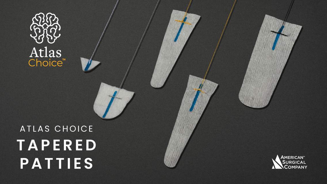
Atlas Choice Tapered Pattie Collection
Low-profile for maximal visualization and protection
Tapered shape designed for retractorless surgery
Unparalleled flexibility and non-stick features
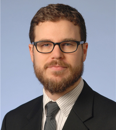
Aaron P. Kamer, MD
Department of Radiology and Imaging Sciences
Neuroradiology Fellowship Director
Indiana University School of Medicine
CONTRIBUTORS
Jacob Eitel, MD
Andrew DeNardo, MD
Sean Dodson, MD
Jordan McDonald, MD
Daniel Murph, MD
Priya Rajagopalan, MD
Daniel Sahlein, MD
John Scott, MD
Rachel Seltman, MD
Medical imaging, in all its forms, is the most powerful tool for noninvasive diagnosis and characterization of preoperative and postoperative neurosurgical diseases. Whether for rapid assessment of intracranial hemorrhage with CT or delineating operability of tumor with MRI, a tremendous amount of information can be gained about your patient’s disease through the proper application of these tools.
Interpreting the spectrum of diseases using the full armamentarium of conventional and advanced imaging techniques requires regular consultation with an experienced radiologist. However, the surgeon with a working knowledge of the imaging principles behind neurosurgical diseases can ensure rapid and effective patient care.
The following volume will describe the working knowledge necessary to handling neurosurgical disorders.
The herculean efforts of Dr. Aaron Kamer and his team (see the list of contributors below) in directing the editing this volume have been unparalleled. Dr. Kamer’s exceptional expertise, inspirational leadership and delightful personality have made this indispensible volume a reality.
Aaron P. Kamer, MD
Department of Clinical Radiology and Imaging Sciences
Residency Director
Indiana University School of Medicine
Contributors
Andrew DeNardo, MD
Sean Dodson, MD
Jordan McDonald, MD
Daniel Murph, MD
Priya Rajagopalan, MD
Daniel Sahlein, MD
John Scott, MD
Rachel Seltman, MD
Please login to post a comment.


Comments: