Internal Cerebral Veins in the Roof of the Third Ventricle
6030
Surgical Correlation
Tags
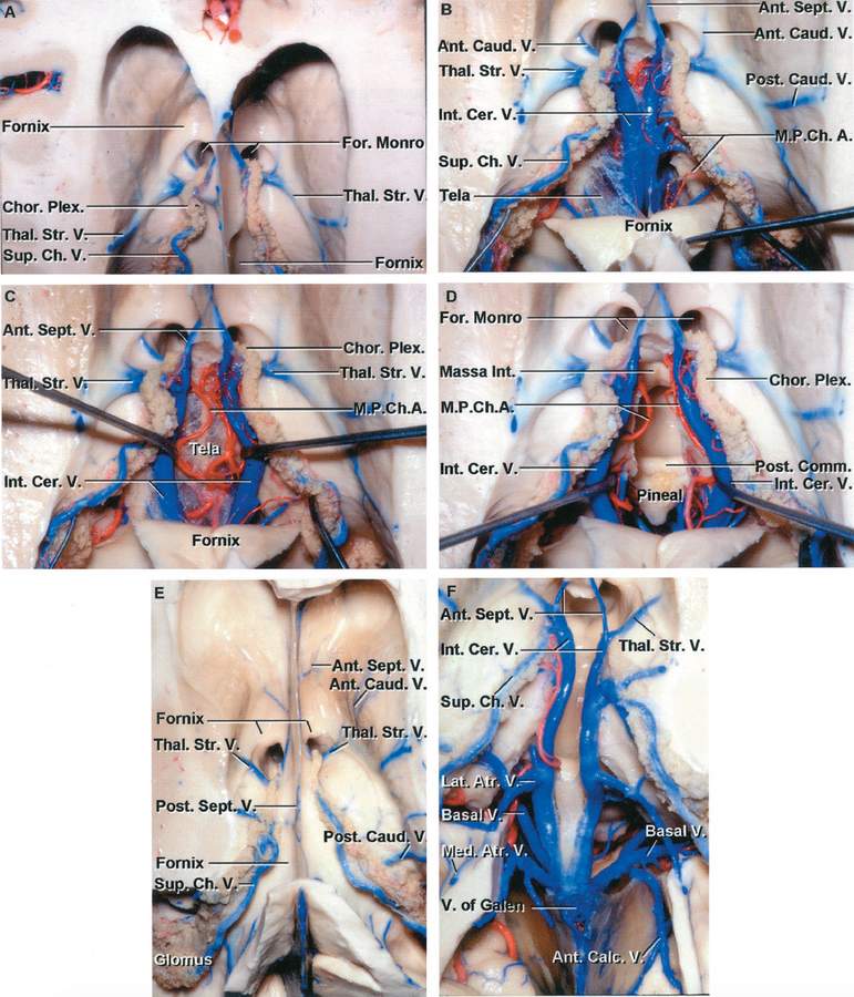
A, Superior view of the frontal horn and body. The thalamostriate and superior choroidal veins converge on the posterior edge of the foramen of Monro. The superior and anterior margin of the foramen of Monro is formed by the fornix. B, The fornix has been folded backward to expose the tela choroidea and the internal cerebral veins in the roof of the third ventricle. A thin layer of ependyma extends above and partially hides the thalamostriate veins coursing along the sulcus between the thalamus and caudate nucleus. The anterior caudate and anterior septal veins cross the lateral and medial wall of the frontal horn. The posterior caudate veins cross the lateral wall of the body of the ventricle. Only a small part of the upper layer of tela located between the fornix and internal cerebral veins remains. C, The internal cerebral veins have been separated to expose the branches of the medial posterior choroidal artery and the lower layer of tela choroidea that forms the floor of the velum interpositum in the roof of the third ventricle. The lower wall of the velum interpositum, in which the internal cerebral veins and medial posterior choroidal arteries course, is formed by the layer of tela attached along the medial side of the thalamus to the striae medullaris thalami. D, The lower layer of tela has been opened and the internal cerebral veins and the medial posterior choroidal arteries have been retracted to expose the posterior commissure, pineal gland, and massa intermedia. E, Another hemisphere. The upper part of the hemisphere has been removed to expose the frontal horn, body and atrium of the lateral ventricle. The choroid plexus is attached along the choroidal fissure. The anterior and posterior caudate veins cross the lateral wall and the anterior and posterior septal veins cross the medial wall of the frontal horn and body of the lateral ventricle. The superior choroidal veins course along the choroid plexus. The thalamostriate veins pass through the posterior margin of the foramen of Monro. The choroid plexus in the atrium expands to a large tuft called the glomus. F, The body of the fornix has been removed to expose the internal cerebral veins coursing in the roof the third ventricle. The medial and lateral atrial and anterior calcarine veins join the posterior end of the internal cerebral veins. The basal veins are exposed below and lateral to the internal cerebral veins. (Images courtesy of AL Rhoton, Jr.)


