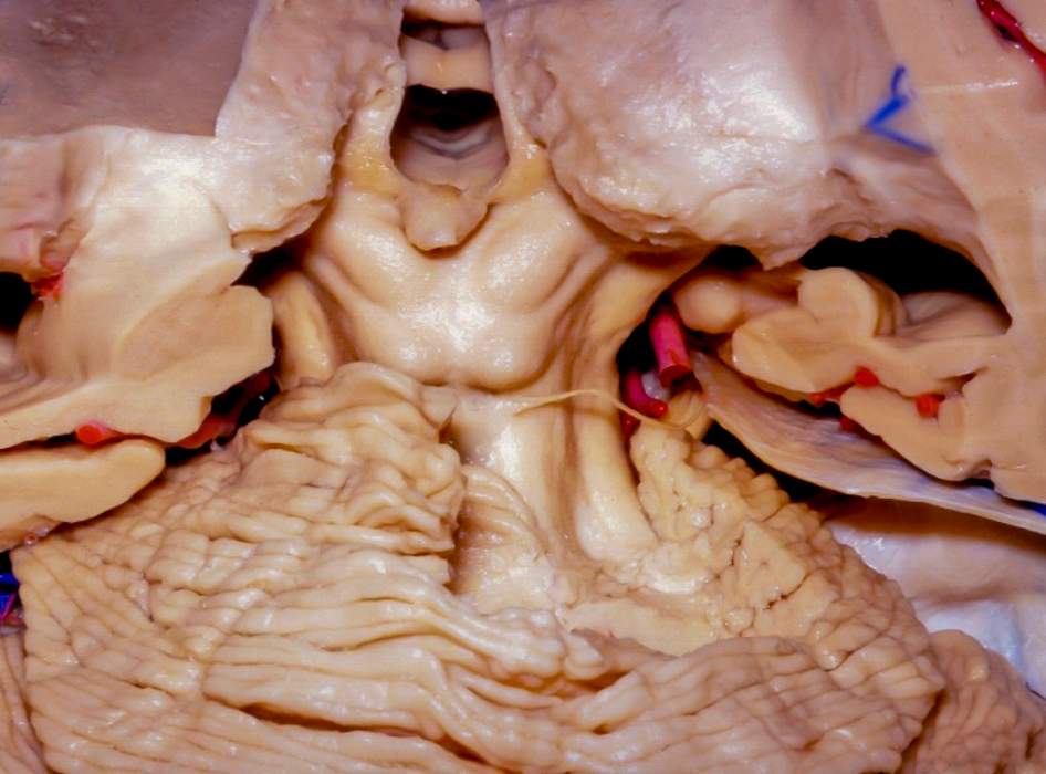
Quadrigeminal Plate and Pineal Region
5438

5438

Dorsal view of the midbrain and quadrigeminal (tectal) plate. Visualized are the superior and inferior colliculi of the midbrain. The trochlear nerve arises from the dorsal midbrain at the level of the cerebellomesencephalic fissure and courses laterally around the midbrain. The pineal gland and third ventricle are visualized after removal of the velum interpositum. (Image courtesy of AL Rhoton, Jr.)
Check to see if you have access through your library or institution.
Start your 30-day free trial or subscribe to access the most comprehensive collection of advanced microneurosurgical techniques. The Neurosurgical Atlas collection presents the nuances of technique for complex cranial and spinal cord operations.
Login or registerCopyright © 2024 Neurosurgical Atlas, Inc. All Rights Reserved. | End User License Agreement | Privacy Policy | Report a Problem
