

Principles of Intraventricular Surgery Free


Figure 1: Walter Dandy demonstrated manual removal of a large ventricular tumor through the parietal transcortical approach (Dandy WE. The Brain. Hagerstown, MD: W.F. Prior Company, 1966.) Dandy pioneered techniques for conquering the surgery of the ventricular system.
Ventricular tumors are rather uncommon, accounting for less than 1% of intracranial lesions. They present a particular technical challenge because of their difficult-to-reach location and the neighboring vital diencephalic and brainstem structures.
Intraventricular tumors represent approximately 10% of all central nervous system neoplasms, and only 10% are exclusively contained within the ventricular system. The tumor is classified based on its intra-axial and extra-axial components.
The most common primary brain tumors include colloid cysts, craniopharyngiomas, astrocytomas, choroid plexus papillomas, ependymomas, epidermoid and dermoid cysts. The most common secondary tumors include meningiomas, gliomas, pituitary adenomas, and arachnoid cysts. These tumors are typically benign, grow at a slow rate, and are usually not identified until they reach a large size, at which point the clinical presentation is a result of either obstructive hydrocephalus or gross compression.
Branches of the choroidal vessels that are reached late in resection supply these masses. These lesions’ characteristics, such as their large size, deep location, and high vascularity, add to the technical complexity of the operation. The indispensable neurologic function associated with the walls of the ventricle and the surrounding deep nuclei should be preserved.

Atlas Choice Tapered Pattie Collection
Low-profile for maximal visualization and protection
Tapered shape designed for retractorless surgery
Unparalleled flexibility and non-stick features
Diagnosis
The mass effect of the tumor results in either obstructive hydrocephalus or a compressive syndrome of deep brain structures. Many patients with these tumors present asymptomatically and the tumors are detected during a diagnostic workup for unrelated headaches.
Patients presenting with large intraventricular masses may suffer from varied neurologic deficits. Common clinical findings may include headaches, poor balance, cognitive dysfunction, personality changes, motor weakness, ataxia, visual disturbances, and seizures as these masses grow and expand into the deep white matter. Patients may be unaware of their memory difficulties and should undergo preoperative neuropsychological testing so that postoperative deficits can be adequately assessed.
Obstructive hydrocephalus can manifest temporally as acute, chronic, or intermittent, as has been described with colloid cysts. Acute obstructive hydrocephalus may present with acute onset headache, nausea, and/or vomiting. Extremely rarely, this mechanism may lead to sudden death with colloid cysts. The intermittent presentation manifests uniquely as posturally-induced nausea, vomiting, headache, or an altered level of consciousness.
Compression syndromes of the surrounding structures compose the other major facet of the clinical presentation. Midbrain compression syndromes commonly seen with intraventricular tumors can present with motor dysfunction, sensory disturbance, or Parinaud’s syndrome.
Parinaud’s syndrome is related to compression of the rostral interstitial nucleus of the medial longitudinal fasciculus within the midbrain tectum by the tumors of the pineal region and tectal plate. Parinaud’s syndrome includes vertical gaze palsy (primarily upward gaze), upbeat or downbeat nystagmus, and impaired convergence.
Many fourth ventricular tumors, including medulloblastomas, often present with compression syndrome of the cerebellar tracts, such as ataxia.
Evaluation
Radiography is the primary mode of evaluation for intraventricular tumors, and magnetic resonance imaging (MRI) is the gold standard. MRI permits a detailed evaluation of the tumor size, vascularity, and extent of invasion. It also permits evaluation of the peritumoral environment, associated edema, and ventricular anatomy, all which are important for preoperative planning.
An in-depth understanding of the three-dimensional anatomy of deep lesions is important for selection of the appropriate operative corridor along the long axis of the mass to minimize brain retraction and transgression. As the lesion’s depth advances, the operative working angles become more important than the simple extent of exposure.
Therefore, reconstructed axial, sagittal, and coronal MRI planes are pertinent for providing the operator with the necessary information for the point of entry into the ventricle. The tumor must be analyzed from multiple angles. Intraventricular lesions typically have a “cap” of cerebrospinal fluid (CSF) surrounding the tumor. As these lesions tend to reach large sizes before their identification, normal anatomy may be distorted and often difficult to identify preoperatively.
Special attention is required to study imperative ventricular veins such as the thalamostriate veins. The functionality of the overlying cortices plays an important role in defining the operative approach. The vascularity of the tumor can increase the risk of operation because the deep narrow surgical corridor hinders efficient management of torrential bleeding.
| Tumor Type | Key MRI Findings | Key CT Findings |
| Colloid cyst |
|
|
| Craniopharyngioma |
|
|
|
Low-grade astrocytoma |
|
|
|
|
|
|
|
|
|
|
|
|
Meningioma |
|
|
|
|
|
|
|
|
|
|
|
|
|
|
| Pineoblastoma |
|
|
| Choroid Plexus Papilloma |
|
|
Indications for Surgery
Most ventricular tumors are asymptomatic and found incidentally. These tumors may be treated expectantly with serial imaging if the initial imaging findings are consistent with a benign or slow-growing tumor (i.e., subependymoma).
Following identification of a symptomatic ventricular tumor, the first-line therapy is complete surgical resection to make a reliable diagnosis, alleviate tumor mass effect, and minimize the risk of future growth. The exception to this management principle is treatment of pineal region masses, which often require either CSF analysis or stereotactic/endoscopic biopsy for further evaluation to rule out a nonoperative germ cell tumor before surgical resection.
The timeframe for surgical intervention is elective because of the slowly progressive nature usually found with these lesions. However, diagnosis of acute intratumoral hemorrhage or acute obstructive hydrocephalus is considered an emergency, and immediate surgical intervention in the form of resection or ventriculostomy is warranted, respectively.
Preoperative Considerations
A number of factors influence the choice of operative corridor. Because some transgression of normal brain is necessary to reach these deep lesions, the selection of the operative corridor plays an important role in the final outcome of the patient. The risks and benefits of each corridor must be carefully and objectively balanced.
I believe the following considerations (listed in the order of importance) should be carefully analyzed before tackling an intraventricular tumor and selecting a safe operative access route:
- minimal transgression and retraction of normal/functional brain,
- expanded working angles to allow effective gross total tumor resection,
- early exposure of vital structures and access to the tumor’s blood supply, and
- the technical difficulty of the operative route.
You can see that the technical complexity of the approach is the least important consideration. Deep seated lesions provide the most opportunity for the operator to be innovative in designing new operative approaches to avoid brain transgression. For example, contralateral interhemispheric approaches provide more flexible angles to the intraventricular tumors located away from the midline under less ipsilateral brain retraction.
The complexity of the approach warrants thorough preoperative radiographic evaluation of the tumor and acquisition of MRI data sufficient for intraoperative navigation. The complex ventricular anatomy, often distorted by the neoplasm, can also be navigated with the assistance of an endoscope. Moreover, the operative reach through narrow operative corridors can be extended using endoscopic-assisted microsurgery.
Tumors that affect the third ventricular wall require preoperative endocrinologic and neuro-ophthalmologic evaluation. Caudal fourth ventricular lesions should have otolaryngologic assessment of the lower cranial nerves’ function.
General Intraoperative Considerations
Choice of patient positioning depends on the chosen operative approach. The supine or lateral position may be used so that the sagittal suture is parallel to the floor. With this head configuration, the falx can hold the contralateral hemisphere out of the surgeon’s way while gravity retraction can expand the interhemispheric corridor.
Resection of lateral ventricular lesions may require longer than average operative times, so all of the pressure points should be well padded during patient positioning on the operating room table.
The location of the bone flap affects access to the ventricle; the superior sagittal sinus is often unroofed for access to the midline lateral ventricular lesions. Hydrocephalus can cause distortion and stretch of the surrounding white matter tracts, making them more susceptible to injury from operative manipulation or retraction. I often use a lumbar or ventricular drainage catheter to decompress the brain during the early stages of the operation (i.e., interhemispheric dissection). The ventricular catheter may be placed opposite the side of operative approach to prevent its interference with the incision and its accidental dislodgement.
For transcortical routes, the cortical incision must be carefully planned to avoid eloquent cortices and their corresponding white matter tracts. Longer but safer pathways to the lesion along with incisions through the sulci and fissures while minimizing cortical retraction all help preserve neurologic function.
Intraoperative electrophysiologic monitoring plays a role in ventricular surgery because of the vulnerability of the periventricular structures, such as the thalamus and brainstem, to operative manipulations. Specific modalities to monitor include motor, somatosensory, and brainstem auditory evoked potentials. A decline in any of these parameters should evoke a rapid response by the surgeon. These responses may include reconsideration of further brain transgression, repositioning of the retractor blade, briskly debulking an acutely expanding intralesional hemorrhagic mass, or CSF drainage.
Because of the long working distance and the need for brain transgression in ventricular surgery, the use of MRI-based navigation is critical for precise localization of the transcortical or transcallosal incisions and successful ventricular navigation. Intraoperative ultrasonography can complement MRI-based navigation in select cases to account for brain shift after ventricular CSF drainage.
The staining of the ventricular system with blood has undesirable ramifications. Therefore, it is important to control the lesion’s vascular supply early in dissection to allow piecemeal resection in deep and narrow but pristine transventricular operative corridors. The rest of the ventricular system should be isolated from the operative field using cotton patties to avoid drainage and entry of blood in other ventricular cavities.
Indiscriminate excessive retraction on the surrounding normal brain and impulsive bipolar coagulation of the ventricular walls are two of the most common causes of new postoperative deficits and must be avoided. These maneuvers often occur in reaction to torrential bleeding when the operator is aimlessly trying to improve visualization in a deep operative corridor and abruptly stop bleeding.
It is important to protect the foramen of Monro when bleeding occurs. Placing a cotton patty at the foramen prevents blood from pooling in the ventricles. All bleeding areas should be identified and controlled prior to closure. This caveat does not mean aggressive coagulation of the walls, but rather patient use of irrigation to achieve hemostasis and clear the field when venous oozing occurs.
Adequate venous drainage is important to prevent venous infarction of the vital diencephalic structures with minimal venous collaterals. There are no clear and reliable principles to determine which veins should be preserved and which can be safely sacrificed. Preoperative angiograms can provide valuable information about the location and dominance of the veins along the operative trajectory and within the ventricles. One of the septal veins may be safely sacrificed if necessary. Thalamostriate and internal cerebral veins are indispensable. I do not believe unilateral sacrifice of a thalamostriate vein is either safe or necessary.
Most ventricular lesions are benign and slow-growing tumors. Subtotal resection may be preferable to complete resection if the lesion involves deep structures such as ventricular walls, the fornices, thalamus, or basal ganglia. If lesions have medial attachments, the surgeon may need to protect the fornix by leaving some tumor behind. Manipulation of the foniceal systems should be minimized because of the high risk of memory dysfunction, even after their minimal injury.
Operative Anatomy
The complexity of the ventricular anatomy demands a dedicated chapter. Please refer to the Anatomy of the Ventricular System chapter for further details.
Overview of Surgical Approaches
To discuss the approaches for intraventricular surgery, I have organized the techniques based on the ventricle of interest. These approaches are briefly described below with an emphasis on the advantages and disadvantages for each. Further in-depth discussions of the individual approaches are found in the respective chapters.
Surgical Approaches to the Lateral Ventricle
The position of the lesion within the lateral ventricle will determine the most appropriate operative trajectory, either anterior, anterolateral, or posterior. This necessitates passing through either the cerebral hemisphere (transcortical) or corpus callosum (transcallosal) to reach the ventricular lumen. Since the working distance is long and the working space is narrow, the importance of flexible working angles cannot be understated. These angles are most practical when the lesion is approached along its long axis.
The superiority of transcortical over transcallosal approaches is highly controversial. The theoretical advantages of the transcortical approach are as follows: the parasagittal veins are not a concern, and tedious interhemispheric arachnoid dissection is not required. However, the projection fibers in the frontal lobe are disrupted. I believe transcortical trajectories (anterior transfrontal versus anterior transcallosal) have less flexible working angles.
Some studies have suggested that the transcortical approach increases the risk of postoperative seizures; however, this finding has not been conclusively confirmed by subsequent studies.
Most importantly, the cortical or callosal incisions should be carefully and strategically planned to lessen the risk of postoperative deficits while providing a sufficiently wide operative passage for exposure and gross total resection of the lateral ventricular lesion. For more discussion of these principles and more extensive details on the relevant approaches, see the Lateral Ventricular Tumors chapter.
|
Lesion Location |
Suggested Approaches |
|
Frontal horn |
|
|
Body |
|
|
Atrium or trigone |
|
|
Temporal horn |
|
|
Occipital horn |
|
Anterior Interhemispheric Transcallosal Approach
Transcallosal approaches are ideal for strictly midline lesions without significant lateral expansion. The anterior interhemispheric transcallosal approach facilitates access to the frontal horn and body of the lateral ventricle, in addition to exposure of the third ventricle. Extensive lateral expansion of the tumor on the ipsilateral side, away from the midline, within the frontal horn is a contraindication to the use of this approach. Transcortical or contralateral transcallosal trajectories are often more appropriate. Bilateral tumor extensions away from the midline indicate the need for the intraparietal transsulcal approach to expose the lesion along its long axis.
The surgeon should avoid injuring the genu of the internal capsule, located lateral to the foramen of Monro and separated by a thin layer of the ventricular capsule. This location is also significant because it is where the thalamostriate vein orients medially to anastomose with the internal cerebral vein.

Figure 2: The basic principles of the anterior interhemispheric transcallosal approach are illustrated. Despite the presence of hydrocephalus, I use lumbar drainage to minimize brain retraction during interhemispheric dissection. The anteroposterior location of the craniotomy may be slightly adjusted based on the location of the tumor within the ventricle. The reach of this approach is shaded in green (inset image). Lateral position allows the use of gravity retraction.
Transfrontal Transcortical Approach (via Middle Frontal Gyrus)
The anterior transfrontal transcortical approach is an acceptable technique for resection of lesions that are mainly limited to and filling the frontal horn. This technique involves a transcortical approach through the middle frontal gyrus. It is ideal for patients with large ventricles and nondominant lesions.
Compared with the transcallosal approach, the anterior transcortical approach potentially spares the parasagittal veins and interhemispheric dissection, but it disrupts some of the frontal commissural and projection fibers.

Figure 3: The transfrontal transcortical approach provides exposure of the frontal horn and the foramen of Monro. The posterior and lateral limits of this approach are restrictive and best suited for lesions predominantly residing in the frontal horn and the foramen of Monro.
Posterior Interhemispheric Transcallosal Approach
This approach is designed to reach the posterior body of the ventricle. This route can also be effective for exposing large pineal region masses with supratentorial extension. The splenium is spared, and only a small section of the posterior aspect of the corpus callosum is transected.

Figure 4: The posterior interhemispheric transcallosal approach exploits a callosotomy just anterior to the splenium that must be preserved. This route also exposes posterior third ventricular and large pineal region tumors with cranial extension.
Transparietal Transcortical Approach (via Superior or Inferior Parietal Lobule)
The transparietal transcortical route via the superior parietal lobe is used by many of my colleagues for exposing posterior ventricular lesions. The variant of this approach through the intraparietal sulcus is used to reach the trigone or atrium. The disadvantages of both approaches are the neurologic deficits that can result from dissection of the underlying white matter fibers.
Approaching through the dominant superior or inferior (temporoparietal junction) parietal lobule can result in dyslexia, agraphia, acalculia, visual field deficits, and finger agnosia due to retracting or traversing the angular gyrus. In the nondominant hemisphere, the same approach can result in impaired memory of visual-spatial information, neglect, and visual field deficits.
I prefer the contralateral interhemispheric transfalcine transprecuneus approach for accessing the periatrial and peritrigonal regions.
Contralateral Interhemispheric Transfalcine Transprecuneus Approach
Please refer to the chapter on the contralateral interhemispheric transfalcine transprecuneus approach for more information. This is a very flexible route for large lesions within the atrium and affecting the medial wall of the periatrium.
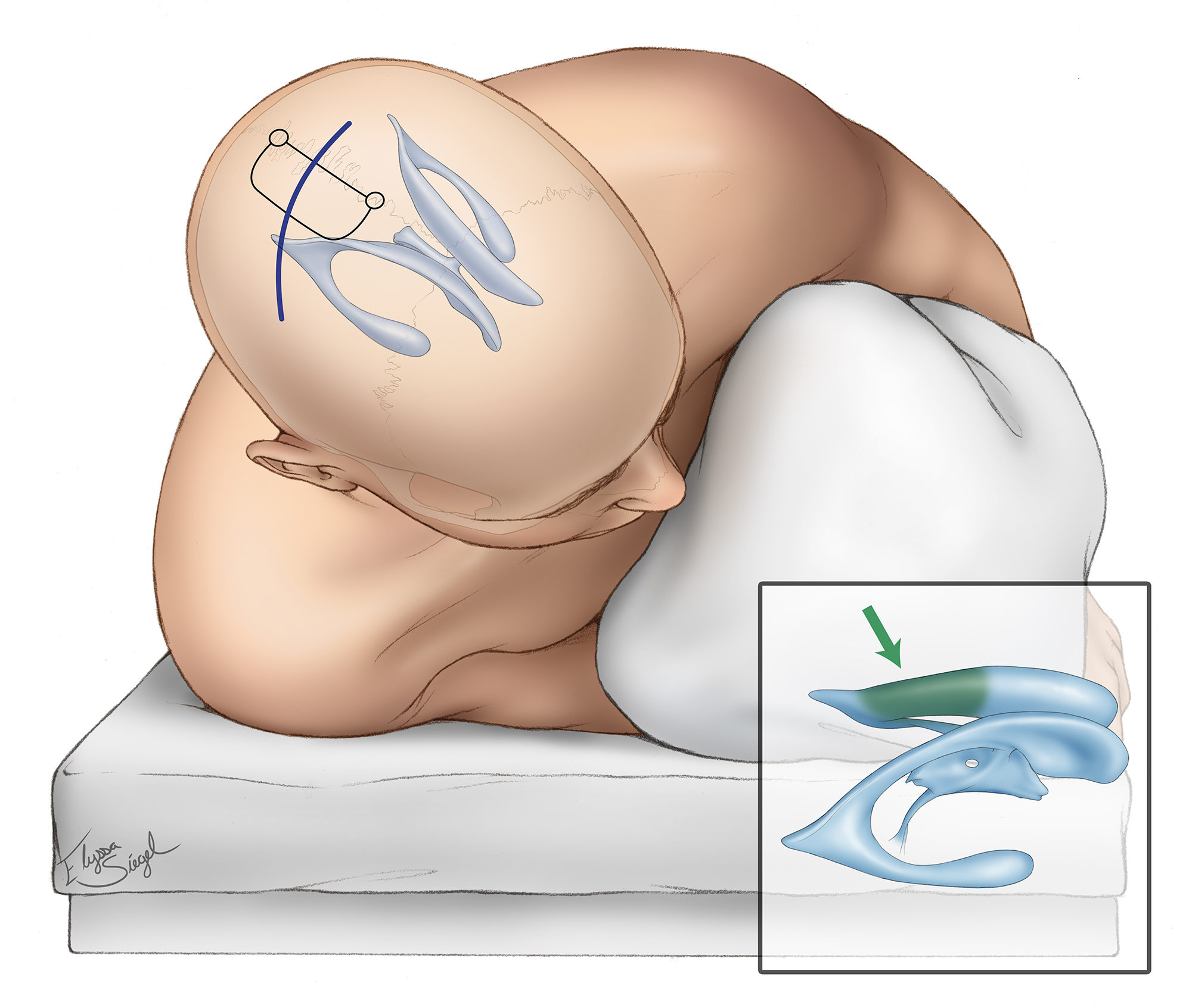
Figure 5: The contralateral interhemispheric transfalcine transprecuneus approach is ideal for periatrial lesions, including arteriovenous malformations along the medial wall of the atrium.
Posterior Ipsilateral Interhemispheric Transcingulate/Transprecuneus Approach
The posterior interhemispheric transcingulate/transprecuneus approach is a reasonable option for lesions within the medial wall of the atrium and occipital horn. The advantage of this approach is that the surgeon is able to avoid contact with the posterior segment of the optic radiation along the lateral ventricular capsule of the atrium and occipital horn.
The lateral reach of this trajectory is limited because of the risk of retraction injury and presence of tethering parasagittal veins.
Anterior Temporal Neocortical Resection
This approach is one of my favorite routes for reaching the anterior and middle temporal horn lesions (anterior to the plane of the cerebral peduncle). Very limited anterior temporal resection (~2.5–3cm) is well tolerated in dominant and nondominant hemispheres and provides excellent exposure.
Other variations of the transcortical approaches to the temporal horn (see below) lead to more extensive cortical transgression around the midsection of the lobe where language function is more prevalent.

Figure 6: Anterior temporal neocortical resection can provide the necessary exposure for the anterior part of the temporal horn, amygdala, uncus and hippocampus. The oblique or tangential operative trajectory can even reach the mid to posterior part of the horn and associated structures.
Transtemporal Transcortical Approach (via Middle Temporal Gyrus)
The transtemporal transcortical pathway via the middle temporal gyrus corticotomy has been employed for reaching the midsection of the temporal horn and hippocampus. Disadvantages of this approach relate to the neurodeficits resulting from the sacrifice of the middle part of the middle and inferior temporal gyri. Although a small corticotomy is initially made, by the time the resection is complete more extensive retraction injury to the surrounding cortices is inflicted.
Approaching through the middle temporal gyrus is high-risk due to variations of the language map in the dominant hemisphere, requiring cortical stimulation mapping for reliable localization of function. An approach through the nondominant hemisphere avoids this risk.
The limited anterior temporal lobe resection (see above) provides an adequate but oblique and tangential working angle to the mid portion of the temporal horn. I have witnessed minimal to no untoward effects using this approach.

Figure 7: The transtemporal approach through the dominant middle temporal gyrus can endanger language. It may be used safely for the nondominant lobe.
Transtemporal Transsulcal Approach (via Occipitotemporal Sulcus)
The occipitotemporal transsulcus approach is useful for lesions located posteriorly within the temporal horn. The major disadvantage of this approach is the risk of postoperative visual field deficits, most commonly a superior quadrant field deficit. This approach is not ideal, especially on the dominant hemisphere. Therefore benefits and risk of this approach must be considered judiciously.
I do not use this approach. I prefer the use of small anterior temporal neocortical resection and a tangential operative corridor to the middle portion of the temporal horn. For more posteriorly situated lesions, around the area of the Calcar avis, I use the supracerebellar transtentorial transparahippocampal trajectory.
Transsylvian Approach
The transsylvian approach can also be used for accessing the anterior temporal horn. Historically, this approach was implemented for amygdalohippocampectomies. For more details see selective amygdalohippocampectomy.
The major advantage of this approach pertains to the dominant amygdala, anterior hippocampal, and parahippocampal limbic tumors that extend into the temporal horn. For these lesions, the transsylvian approach permits resection without transgression of the dominant neocortex of the superior, middle, and inferior temporal gyri.
I use this approach sparingly for small dominant amygdalar and anterior hippocampal lesions. This approach is challenging because the middle cerebral artery branches must be mobilized and manipulated within the fissure with the attendant risks of vascular injury or spasm. In addition, the working corridor is limited to the anterior temporal horn and is less flexible for lesions extending to the midsection of the horn. Overall, there are limited indications for the use of this corridor.

Figure 8: The transsylvian corridor is limited and places the middle cerebral artery branches at risk. This sketch demonstrates the resection of the right amygdala and anterior hippocampus through this route. The oculomotor nerve is apparent.
Posterior Interhemispheric Transcortical Approach to the Occipital Horn
This interhemispheric approach provides a pathway toward the occipital horn. Most patients suffer from preoperative visual deficits that allow a transcortical route through the medial occipital lobe without causing new neurologic complications.

Figure 9: The interhemispheric trancortical approach to the occipital horn is a reasonable trajectory for reaching the horn among the patients who are suffering from irreversible visual dysfunction.
Occipital Neocortical Resection
This transcortical approach has a high likelihood of generating a permanent homonymous visual field deficit. Therefore this approach is only appropriate for patients with an irreversible pre-operative homonymous deficit without the likelihood of reversal.
Surgical Approaches to the Third Ventricle
Third ventricular lesions are among the most challenging lesions to reach and resect safely and effectively. The deep midline position of these tumors situates them adjacent to multiple vital structures including the pituitary stalk, optic chiasm, thalamus, hypothalamus and internal cerebral veins.
Surgery of this region requires pristine familiarity with the anatomy and scrupulous planning and surgical technique. For further discussion of surgery within the third chamber, please see the chapters on the Anterior and Posterior Third Ventricular Tumors.
| Third Ventricular Approaches | |
|
Lesion Location |
Suggested Approaches |
| Anterior |
|
|
Parasellar |
|
|
Lateral ventricle extension |
|
|
Posterior |
|
|
Pineal Region |
|
Anterior Interhemispheric Transcallosal Expanded Transforaminal Transvenous Transchoroidal Approach
This is my favorite approach for reaching anterior and middle third ventricular lesions. The trancallosal approach exposes the ipsilateral lateral ventricle. Next, the foramen of Monroe is enlarged through transection of the septal vein and its disconnection from the thalamostriate vein.
This maneuver allows enlargement of the foramen through a minimal anterior transchoroidal dissection without significant manipulation of the thalamus or thalamostriate vein. This approach is not ideal for very posterior lesions including the ones affecting the pineal region. Please refer to the corresponding chapter on Anterior Third Ventricular Tumors.

Figure 10: The transection of the anterior septal vein and transchoroidal dissection around the thalamostriate vein provides an excellent exposure of the anterior and middle portions of the third ventricle.
Anterior Transfrontal Transcortical Transforaminal Approach
This is a reasonable approach for reaching most anterior third ventricular lesions. For further details, please see the Colloid Cyst (Trasncortical Approach) chapter.
Compared to its transcallosal counterpart, its exposure is somewhat limited in its anteroposterior and lateral-medial working angles and is appropriate for smaller lesions such as colloid cysts. Asymmetric tumors can be tackled through a contralateral transforaminal trajectory (“cross-court route”) to reach the lateral aspect of the tumor.
The anterior transcortical approach also requires transection of some of the commissural and projection fibers. However, it spares the callosum.

Figure 11: The transcortical transforaminal approach can offer a similar exposure to the one provided by its transcallosal counterpart. However, the exposure is more limited for manipulation of large lesions extending beyond the anterior third ventricle. Note entry into the left lateral ventricle.
Subfrontal Translamina Terminalis Approach
This approach is a reasonable one for lesions that are within the anterior segment of the third ventricle, particularly those anterior to a plane running from the cerebral aqueduct to the anterior margin of the foramen of Monro. It is especially favorable if the tumor is extending through the lamina terminalis and has a smaller suprasellar component. For further details, please refer to the chapter on the subfrontal translamina terminalis approach.
Tumors with primarily suprasellar components are resected through the endoscopic transnasal transsphenoidal translamina terminalis approach. Please see the chapter on craniopharyngioma.
This approach offers the advantage of providing excellent visualization of the lamina terminalis, optic nerves, optic chiasm, bilateral internal carotid arteries, anterior communicating artery complex, bilateral A2 segments, posterior communicating arteries, corresponding perforating branches, and the pituitary stalk.
However, its reach for tumors that do not enlarge the lamina terminalis is limited as the safe working space and angles through the non-enlarged lamina terminalis is nonflexible and restricted. Gross total tumor resection can be problematic. The anterior interhemispheric variation of this approach may provide more extended operative space.

Figure 12: The subfrontal translamina terminalis approach provides limited exposure to the anterior third ventricle and is suitable for lesions enlarging the lamina terminalis and its operative pathway.
Endoscopic Transnasal Transsphenoidal Translamina Terminalis Approach
This approach is very useful and favorable approach for resection of parasellar lesions with third ventricular extension. These tumors are often a craniopharyngioma, pituitary adenoma, or Rathke’s cleft cyst. For further technical details, please refer to the chapter on craniopharyngioma.
The major disadvantage of this approach is the difficulty in performing sufficient microdissection for tumors of dense consistency and adherence to the third ventricular wall, optic structures or perforating vessels. These limitations have been recently overcome with the use of endoscopic microsurgical instruments.

Figure 13: The endoscopic transnasal transsphenoidal translamina terminalis approach provides excellent exposure of third ventricular tumors that have enlarged the lamina terminalis. This corridor is parallel to the long axis of the tumor and allows effective gross total tumor resection through a minimally invasive pathway.
Posterior Interhemispheric Transcallosal Transvenous/Paravenous Variant
Posterior interhemispheric transcallosal transvenous/paravenous variant is appropriate for lesions primarily within the posterior third ventricle, quadrigeminal plate and pineal region.
It provides a unique and suitable surgical field for the area around the posterior internal cerebral veins, straight sinus, vein of Galen, and pineal region. Parasagittal veins are more indispensable in the posterior frontal region; their sacrifice and manipulation can predispose to venous infarction.
Other disadvantages to this approach include injury to the posterior corpus callosum fibers. Despite preservation of the splenium, the risk of auditory deficits, amnesia, mutism, and dyslexia still exists. After transection of the corpus callosum, I work between (intervenous) or around (paravenous) internal cerebral veins. These veins diverge within the posterior roof of the third ventricle and are often further displaced away from each other by the tumor.
Extraocular movement dysfunction ranging in severity up to complete temporary Parinaud’s syndrome, can result from injury of the quadrigeminal plate during this approach. Other sequela of the procedure includes seizures and hemiparesis, most temporary. For further details, please refer to the chapter on Posterior Third Ventricular Tumors.

Figure 14: The posterior interhemispheric transcallosal intervenous variant is a practical route for exposing the posterior third ventricle. The columns of fornix have diverged away from each other at this location and are not usually at risk.
Supracerebellar Transventricular Approach
The infratentorial supracerebellar approach has great utility for small posterior third ventricular lesions, such as cavernous malformations, even if the lesion is not primarily within the pineal region. The advantages of this approach are substantial and include minimal injury to the adjacent structures and conservation of the corpus callosum. The pulvinar can be gently manipulated without significant effects. For further details, refer to the Supracerebellar Transventricular Approach chapter.
The disadvantages of this approach include the vulnerability of the habenula, vein of Galen, and quadrigeminal plate. If damaged, the sequela can include permanent mutism and cognitive impairment.

Figure 15: Small posterior third ventricular tumors can be exposed through the supracerebellar route. A careful study of preoperative imaging is necessary to evaluate the location of the deep veins (vein of Galen, etc) and the feasibility of this approach.
Occipital Interhemispheric Transtentorial Approach
The occipital interhemispheric transtentorial approach is designed to reach posterior third ventricular and pineal region lesions. The disadvantages of this approach include the vulnerability of the occipital lobe, straight sinus and its anastomosing veins, and the splenium.
The injury to these structures consists of cognitive impairment, visual field deficits, and in rare instances split brain syndrome. I have not used this approach because of the real risk of above complications.
Anterior Interhemispheric Transcallosal Interforniceal Approach
This approach has utility for lesions within the anterior to mid segment of the third ventricle. The disadvantages of this approach are the potential for disruption of forniceal projection fibers. However, some tumors erode through or attenuate the forniceal raphe and make this approach ideal. In addition, the presence of cavum septum pellucidum can be helpful. For further details, refer to the Transcallosal Interforniceal Approach chapter.
To avoid injury to the forniceal fibers, the surgeon must be cognizant of retraction force on ventricular, paraventricular, and midline structures. Consequences of forniceal fiber injury include transient and/or permanent amnesia and cognitive impairment.
Surgical Approaches to the Fourth Ventricle
Fourth ventricular tumors pose a surgical challenge, albeit less than their third ventricular counterparts, due to their relation to the brainstem; this relation can vary from simply displacement to invasion.
This challenge is further complicated by the tumor often being concealed by key cerebellar structures including the cerebellar tonsils, cerebellar hemispheres, and/or the vermis. Tumors can involve remote spaces, through extension within the foramen of Luschka, such as the premedullary, cerebellomedullary, prepontine, and anterior spinal cisterns.
These extensions can facilitate involvement of the posterior circulation branches and perforators, including the cranial nerves. The operator must be cognizant of these structures and their exact location during fourth ventricular surgery. For further discussion of these principles, see the Fourth Ventricular Tumors chapter.
Historically, the approach to the fourth ventricular tumors involved either cerebellar hemisphere resection or vermian split. The latter has a real risk of vermian split syndrome, which is characterized by neurobehavioral abnormalities, imbalance and cerebellar mutism. In an attempt to avoid these complications, an alternative approach (telovelar route) has been designed.
Telovelar or transcerebellomedullary Fissure Approach
This approach serves as an alternative to the historical transcerebellar or tansvermian approaches. It facilitates generous exposure of the fourth ventricular space with significantly less disruption of normal structures. For further details, please refer to the chapter on Telovelar Approach in the Cranial Approaches volume.

Figure 16: The telovelar approach provides the least invasive and most effective route to the fourth ventricle.
For large fourth ventricular tumors with significant cranial extension around the superior medullary velum and the pineal region, a small inferior vermian split is safe. Alternatively, a combined telovelar and supracerebellar approach can be attempted.
Closure
Generous irrigation of the ventricular system is imperative to prevent ventricular obstruction postoperatively. Small pieces of hemostatic material (i.e., SURGICEL or Gelfoam powder) cannot be left behind; their removal will minimize the risk of acute postoperative hydrocephalus. Fenestration of the septum pellucidum and lamina terminalis (in select cases) is advised, and may obviate a need for postoperative permanent CSF diversion.
Cerebral spinal fluid should be replaced with warm irrigation at the conclusion of the case to prevent hemispheric collapse and development of subdural fluid collections.
Postoperative Considerations
Patients should be closely monitored postoperatively for acute development of hemorrhage, hydrocephalus, and subdural hematomas. In select patients, a catheter should be left in the ventricle to monitor intracranial pressure and drain intraventricular debris related to surgery.
Within 24 hours of the procedure, a CT scan should be obtained to evaluate for hemorrhage, ventricular size, asymptomatic ischemia or edema and any obvious residual lesion. The patient should be mobilized by day three and the intraventricular catheter removed. The catheter should be removed as soon as possible to prevent infection and ventriculitis.
Intracranial pressure should be kept between 10 and 15 mmHg with the patients’ head elevated at 20 to 30 degrees. Judicious CSF drainage of small amounts is best to remove debris, maintain appropriate pressures and prevent subdural fluid accumulation.
Some patients may require permanent CSF diversion for postoperative hydrocephalus. Permanent shunting may improve speech, motor function, and cognitive abilities in these patients. Further, if ventricular obstruction was not cleared intraoperatively, a shunt may be necessary.
Postoperative deficits vary depending on the approach, size, location, extent of resection and most importantly manipulation of ventricular walls and adjacent normal structures. Visual field loss is one of the most common deficits, occurring in 20-64% of cases.
Cognitive dysfunction and hemiparesis have been reported in 8-30% of patients and frequently occur early in the postoperative period but significantly improve overtime in the majority of patients. There are also possibilities of speech disturbances, particularly with mass lesions of the dominant ventricle.
Postoperative seizures are common with resection regardless of the procedure. Incidence of seizures ranges from 29% to 70%, especially for transcortical routes. Most patients present with hydrocephalus, and despite gross total resection of their tumors, up to 33% require permanent shunting.
Contributor: Benjamin K. Hendricks, MD
Please login to post a comment.


