

Coccidioidomycosis

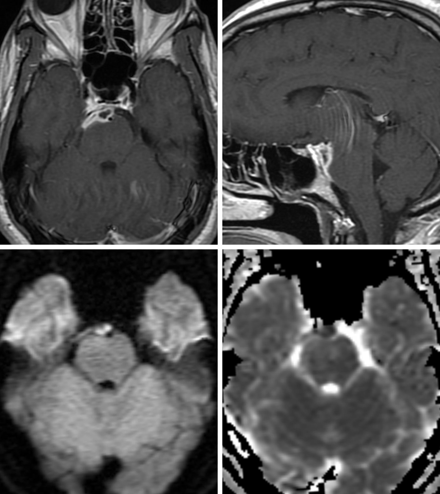
Figure 1: Peripherally enhancing lesion along the ventral pons (top left and top right) demonstrating central restricted diffusion on DWI (bottom left) and ADC (bottom right) images, a finding seen more typically in infectious abscesses than in tumors. When along the surface of the brain, as in this case, coccidioidomycosis can mimic leptomeningeal carcinomatosis.

Figure 2: Axial (top left and top right) and coronal (bottom left) contrast-enhanced T1-weighted images demonstrating numerous tiny enhancing nodules along the basal cisterns and within and along the cerebellum reflecting leptomeningeal disease and occasional parenchymal abscesses, which are common with coccidiomycosis and can mimic small metastases. (Bottom Right) Axial FLAIR image showing hyperintense local edema associated with small parenchymal cerebellar abscesses.

Figure 3: Axial T1-weighted postcontrast images demonstrating prepontine cistern and medial left temporal enhancing coccidiomycosis involvement (top left) and enhancing infectious tissue along the medial anterior left lobe, in the right posterior orbital gyrus and in the left basal ganglia (top right), which can also be seen in dura-based and parenchymal metastatic disease. The parenchymal lesions also demonstrate nonspecific surrounding FLAIR hyperintense edema (bottom left and bottom right) that would also be typical of parenchymal metastatic disease.
Description
- Coccidioides immitis is a dimorphic fungus endemic to the southwestern United States and regions of Mexico and Central and South America
- Primary infection occurs in the lungs after inhalation of airborne arthrospores
- Up to 5% of patients develop disseminated disease, with CNS involvement being the most frequent and severe manifestation

Atlas Choice Tapered Pattie Collection
Low-profile for maximal visualization and protection
Tapered shape designed for retractorless surgery
Unparalleled flexibility and non-stick features
Pathology
- Mycelial form known to grow in the soil and produces spores that become airborne with disturbance of the soil
- Inhalation of the spores leads to pulmonary infection
Clinical Features
- Signs and symptoms
- Headache, nausea, vomiting, and altered mental status with concurrent pulmonary symptoms, septic shock, and fungemia
- Age and gender
- No age or gender predilection
- Prognosis
- Mean survival time
- Without treatment, 4 months
- With treatment (amphotericin B), 21 months
- Mean survival time
Imaging
- General features
- Most common presentation is meningitis with a propensity for the basal cisterns
- Hydrocephalus, infarctions, vasculitis, parenchymal or parameningeal masses and abscesses, periventricular white matter abnormalities and spinal arachnoiditis
- Important to perform spinal imaging, because spinal involvement is common and can lead to change in management
- Modality specific
- CT
- Ill-defined areas of hypoattenuation with intermixed hemorrhage
- Hydrocephalus, herniation
- MRI
- T1WI
- Ill-defined hypointense regions
- T2WI
- Ill-defined hyperintense regions with possible hypointense rim
- DWI
- Restriction along the wall of the abscess
- SWI
- Hypointense signal in the setting of hemorrhage
- Contrast
- Meningeal enhancement in the setting of meningitis
- If cerebral parenchymal involvement, there will be variable enhancement
- T1WI
- CT
- Imaging recommendations
- Standard protocol MRI (including DWI) with intravenous contrast
- Mimic
- Similar to other infectious processes, when walled off, it is difficult to distinguish from other ring-enhancing lesions. However, when presented with basilar cistern involvement, coccidioidomycosis should be included in the differential with tuberculosis and other granulomatous processes such as sarcoidosis
For more information, please see the corresponding chapter in Radiopaedia.
Contributor: Sean Dodson, MD
References
Gupta NA, Iv M, Pandit RP, et al. Imaging manifestations of primary and disseminated coccidioidomycosis. Appl Radiol 2015;February:9–21.
Lammering JC, Iv M, Gupta N, et al. Imaging spectrum of CNS coccidioidomycosis: prevalence and significance of concurrent brain and spinal disease. AJR Am J Roentgenol 2013;200:1334–1346. doi.org/10.2214/AJR.12.9264
Starkey J, Moritani T, Kirby P. MRI of CNS fungal infections: review of aspergillosis to histoplasmosis and everything in between. Clin Neuroradiol 2014;24:217–230. doi.org/10.1007/s00062-014-0305-7
Wrobel CJ, et al. MR findings in acute and chronic coccidioidomycosis meningitis. AJNR Am J Neuroradiol 1992;13:1241–1245.
Please login to post a comment.


