Pituitary Macroadenoma
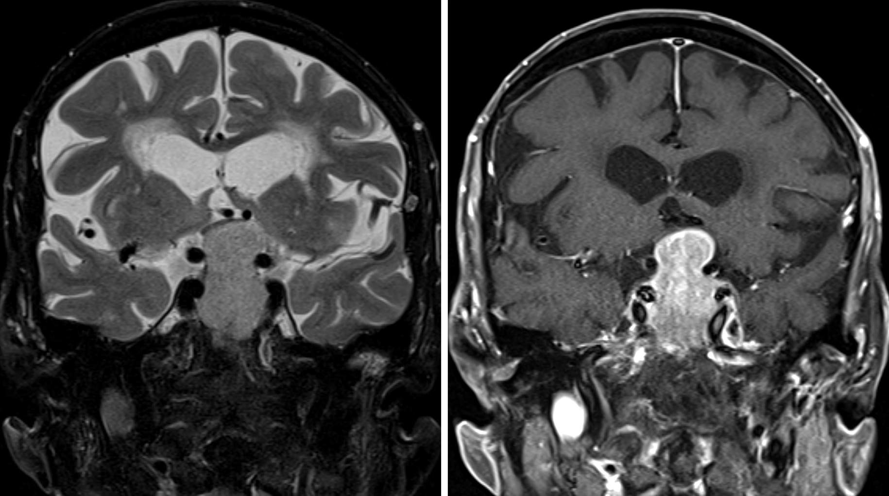
Figure 1: Coronal images through the sella demonstrate a large, short T1 inversion recovery (STIR) hyperintense (left), enhancing (right) mass. The mass has a typical “snowman” shape, with its waist partially constricted by the diaphragma sella. The low-signal-intensity round flow voids of the intracranial internal carotid artery are abutted by this macroadenoma, but lack of significant extension of the mass between the cavernous and supraclinoid segments on either side makes cavernous sinus invasion by the mass less likely. The optic chiasm is deviated superiorly and to the patient’s right.
BASIC DESCRIPTION
- Primary tumor of the adenohypophysis of various cell line differentiations, >10 mm in size
PATHOLOGY
- Low-grade tumors, WHO grade I
- Well differentiated, slow growing
- May secrete hormones according to the cell of origin, most commonly prolactin
CLINICAL FEATURES
- 75% of patients have symptoms related to hormone secretion (secretory)
- Prolactinoma most common
- Women: irregular menses, amenorrhea, galactorrhea
- Men: hypogonadism, decreased libido
- Growth hormone secreting (acromegaly)
- Prolactinoma most common
- Adrenocorticotropic hormone (ACTH) secreting (Cushing disease)
- Thyroid-stimulating hormone (TSH) secreting (hyperthyroidism)
- 25% present with visual symptoms caused by mass effect on the chiasm or optic nerves
- More common in syndromes such as multiple endocrine neoplasia type 1 (MEN1), McCune-Albright, and others
- Pituitary apoplexy is a rare but serious complication requiring emergent surgical attention if acute visual field deficits are apparent
IMAGING
- General
- Sellar mass extending into the suprasellar cistern, displacing the diaphragma sella, often giving a snowman-shaped appearance
- Can have cystic, proteinaceous, or hemorrhagic components
- Can invade the cavernous sinus
- Can invade the underlying sphenoid bone and significantly alter surgical landmarks during transsphenoidal surgery
- CT
- Variable-density enhancing mass obscuring the suprasellar cistern
- Hemorrhage and cystic changes will be hyperdense and hypodense, respectively
- Bony destruction of the sphenoid bone or clivus is often present
- CTA
- Can help delineate position of the surrounding vasculature in large macroadenomas for surgical planning
- MRI
- Coronal images are most helpful for characterizing the morphology of the tumor and its association with neurovascular structures
- Extent of tumor into the “intercarotid interval” determines the likelihood of cavernous sinus invasion
- Comparison of multiple sequences often necessary for identifying the relationship of the tumor to the optic nerves and chiasm
- T1WI: usually isointense to brain/soft tissue; hemorrhagic components can be hyperintense
- T2WI: variable, hyperintense if cystic
- FLAIR: variable, hyperintense if cystic
- DWI: usually intermediate signal
- GRE/SWI: dark signal blooming if hemorrhagic components are present
- T1WI+C: brightly enhancing mass
- MRA
- Can help delineate position of surrounding vasculature in large macroadenomas for surgical planning
- Conventional Angiography
- Inferior petrosal sinus sampling (IPSS) can determine whether a tumor is ACTH secreting
IMAGING RECOMMENDATIONS
- MRI without and with contrast with detailed imaging of the sella
- Thin-cut CT imaging of the sinuses or sella can be helpful for delineating sphenoid sinus anatomy before transsphenoidal resection and during intraoperative navigation
For more information, please see the corresponding chapter in Radiopaedia.
Contributor: Rachel Seltman, MD
References
Aron DC, Tyrrell JB, Wilson CB. Pituitary tumors. Current concepts in diagnosis and management. West J Med 1995;162:340–352.
Chen CC, Carter BS, Wang R, et al. Congress of Neurological Surgeons systematic review and evidence-based guideline on preoperative imaging assessment of patients with suspected nonfunctioning pituitary adenomas. Neurosurgery 2016;79:E524–E526. doi.org/10.1227/NEU.0000000000001391.
Osborn AG. Osborn's Brain: Imaging, Pathology, and Anatomy. Amirsys, Salt Lake City, UT; 2013:699–705.
Raff H. Cushing syndrome: update on testing. Endocrinol Metab Clin North Am 2015;44:43–50. doi.org/10.1016/j.ecl.2014.10.005.
Vance M. Pituitary adenoma: a clinician's perspective. Endocr Pract 2008;14:757–763. doi.org/10.4158/EP.14.6.757.
Please login to post a comment.


