

Cerebral Amyloid Angiopathy/Amyloidoma

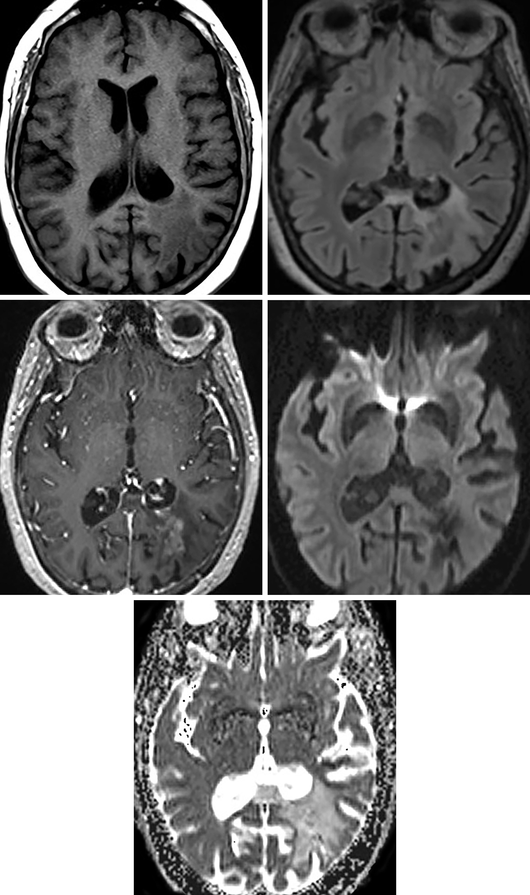
Figure 1: T1-hypointense (top left) and FLAIR-hyperintense (top right) signal within the left parietal white matter radiating medially to the ventricular wall with irregular, somewhat serpiginous enhancement (middle left). (Middle Right and Bottom) No reduction in diffusivity on DWI/ADC images can be seen.
Description
- Cerebrovascular disorder characterized by the deposition of amyloid protein in the media and adventitia of small- and medium-sized vessels of the cerebral cortex, subcortical white matter, and leptomeninges

ATLAS Choice Bipolar Forceps
Designed for your every surgical maneuver
Five tip sizes for brain and spine procedures
Unparalleled non-stick and low-profile features
Pathology
- Unclear
- Thought to be related to a benign clonal proliferation of plasma cells
- Three varieties
- Cerebral amyloid angiopathy, amyloidoma, rare inflammatory subtype
Clinical Features
- Symptoms
- Seizures, headache, focal motor signs, and cognitive decline
- Age
- Angiopathy fairly common in the elderly population
- Amyloidoma found in younger patients; mean age, 48 years
- Gender
- Slightly more common in women (8:5 female/male)
- Associations
- 40% of patients with cerebral amyloid angiopathy have Alzheimer disease
Imaging
- General
- Focal nonhemorrhagic masses
- Mass effect is generally minimal/mild
- Might show moderate/striking enhancement
- Often extends medially to lateral ventricular wall with fine radial enhancing margins
- Radiating pattern from the ventricular wall can help with the diagnosis
- Modality specific
- CT
- Hyperdense, enhances
- MRI
- T1
- Variable appearance
- T2
- Heterogeneous, although more commonly hyperintense
- Contrast
- Variable
- T1
- CT
- Mimic
- Classic amyloid angiopathy can mimic treated or hemorrhagic metastases. The less common amyloidoma can mimic a neoplasm with or without hemorrhage. The radiating pattern from the ventricular wall, when present, can help in differentiating, although this is a fairly nonspecific finding
For more information, please see the corresponding chapter in Radiopaedia.
Contributor: Sean Dodson, MD
References
Chao CP, Kotsenas AL, Broderick DF. Cerebral amyloid angiopathy: CT and MR imaging findings. Radiographics 2006;26:1517–1531. doi.org/10.1148/rg.265055090
Galluci M, Caulo M, Splendiani A, et al. Neuroradiological findings in two cases of isolated amyloidoma of the central nervous system. Neuroradiology 2002;44:333–337. doi.org/10.1007/s00234-001-0728-0
Gandhi D, Wee R, Goyal M. CT and MR imaging of intracerebral amyloidoma: case report and review of the literature. AJNR Am J Neuroradiol 2003;24:519–522.
Haacke EM, DelProposto ZS, Chaturvedi S, et al. Imaging cerebral amyloid angiopathy with susceptibility-weighted imaging. AJNR Am J Neuroradiol 2007;28:316–317.
Landau D, Avgeropoulos N, Ma J. Cerebral amyloidoma mimicking intracranial tumor: a case report. J Med Case Rep 2010;20;4:308. doi.org/10.1186/1752-1947-4-308
Please login to post a comment.


