Transchoroidal Approach to the Third Ventricle Directed Along the Forniceal Side of the Choroidal Fissure
6061
Surgical Correlation
Tags
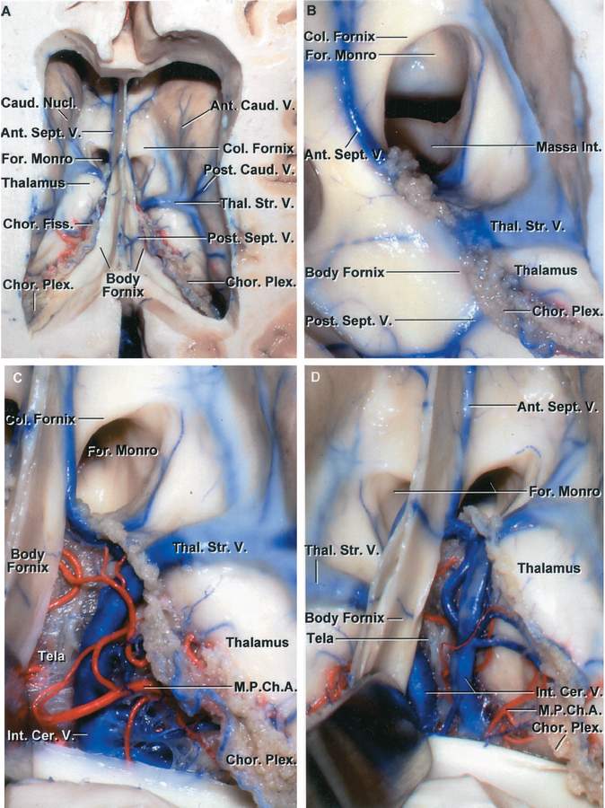
A, Superior view of the frontal horn and body of the lateral ventricle. The body of the fornix forms the upper part of the roof of the third ventricle. The left thalamostriate vein passes through the posterior margin of the foramen of Monro and the right thalamostriate vein passes through the choroidal fissure a few millimeters be-hind the foramen. Anterior septal and anterior caudate veins cross the wall of the frontal horn. Posterior septal and posterior caudate veins cross the wall of the body of the lateral ventricle. The thalamus sits in the floor of the body. The choroidal fissure, located between the thalamus and fornix, is opened by dividing the tenia fornix that attaches the choroid plexus to the lateral edge of the fornix, leaving the attachment of the choroid plexus to the thalamus undisturbed. B, Enlarged view. The columns of the fornix form the anterior and superior wall of the foramen of Monro. The massa intermedia is seen through the foramen. Anterior and posterior septal veins cross the septum pellucidum and fornix. C, The tenia fornix, which attaches the choroid plexus to the fornix, has been divided and the body of the fornix retracted medially to expose the internal cerebral vein and medial posterior choroidal arteries. The lower layer of tela, which attaches to the striae medullaris thalami and forms the floor of the velum interpositum, is intact. D, The separation of the fornix and choroid plexus has been extended posteriorly to the junction of the atrium and body of the ventricle. The lower layer of tela remains intact. (Images courtesy of AL Rhoton, Jr.)


