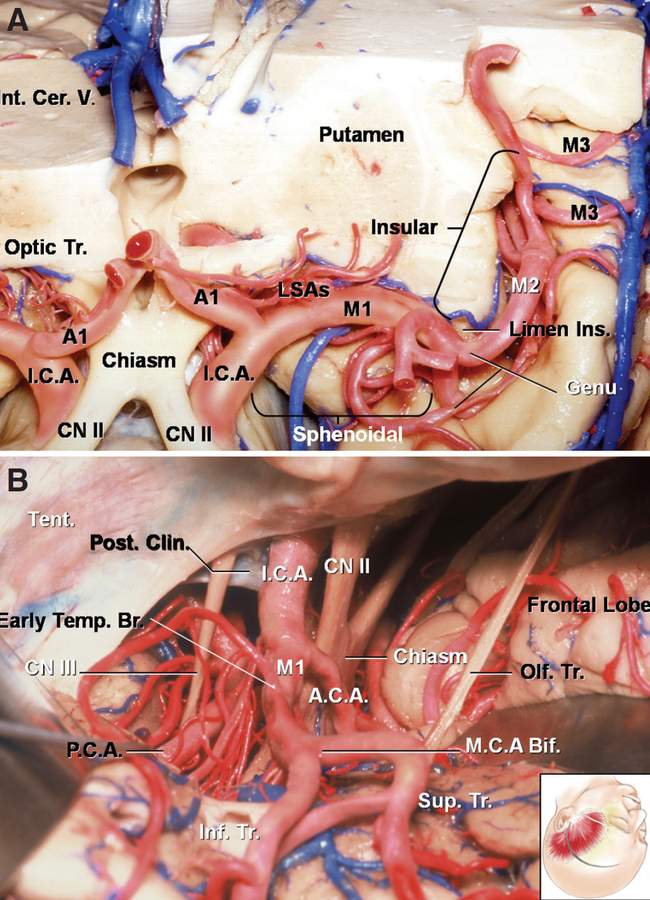
The Middle Cerebral Artery in Coronal and Intraoperative Views
6896
Surgical Correlation
Tags
The Middle Cerebral Artery in Coronal and Intraoperative Views. A, Coronal dissection demonstrating the course of the middle cerebral artery (MCA) within the sphenoidal and insular compartments of the sylvian fissure. The M1 pre- and postbifurcation trunks run within the sphenoidal compartment. At the limen insula, the artery turns to run posteriorly within the insular compartment. The genu marks the division between the M1 and M2 segments of the artery. The M2 arteries give off branches to the lateral cortex, which course over the frontal, parietal, and temporal opercula. The opercular portions of the MCA correspond to the M3 segments. B, Left pterional exposure demonstrating the internal carotid artery (ICA) bifurcation into the anterior cerebral artery (ACA) and MCA. The M1 prebifurcation segment of the MCA extends from the ICA bifurcation to the MCA bifurcation and runs in the sphenoidal compartment of the sylvian fissure. The M1 segment frequently gives off cortical branches before its bifurcation. These branches are early frontal and early temporal arteries. Lenticulostriate arteries (LSAs) often arise from the proximal segment of these early branches. The M1 segment continues a variable distance as M1 postbifurcation trunks before the genu at the level of the limen insula. At the limen insula, the postbifurcation trunks turn to run along the surface of the insula as the M2 segment. The M2 segments run within the insular compartment of the sylvian fissure. The inset shows the patient orientation. (Images courtesy of AL Rhoton, Jr.)


