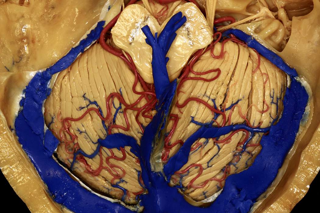
Superior View of the Posterior Fossa after Tentorium Cerebelli Resection
6527

6527

Superior view of the posterior fossa after tentorium cerebelli resection. Notable venous anatomy present along the tentorium includes the superior petrosal sinus, which drains the cavernous sinus to the transverse-sigmoid junction. The transverse sinus is visible in its entirety within the tentorium. The internal cerebral veins and the basal veins form the great vein of Galen, which drains to the straight sinus and then the torcula. The quadrangular lobule (part of the anterior lobe of the cerebellum) and simple lobule (part of the posterior lobe of the cerebellum) are separated by the primary fissure. (Image courtesy of AL Rhoton, Jr.)
Check to see if you have access through your library or institution.
Start your 30-day free trial or subscribe to access the most comprehensive collection of advanced microneurosurgical techniques. The Neurosurgical Atlas collection presents the nuances of technique for complex cranial and spinal cord operations.
Login or registerCopyright © 2024 Neurosurgical Atlas, Inc. All Rights Reserved. | End User License Agreement | Privacy Policy | Report a Problem
