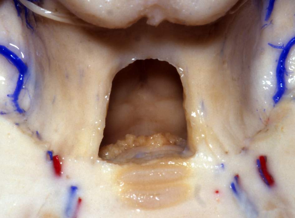
Superior View of the Fourth Ventricle
6318

Surgical Correlation
Tags
Superior view of the fourth ventricle. The superior medullary velum has been removed to reveal the fourth ventricle from above. The superior cerebellar peduncles course within the dorsolateral walls of the rostral fourth ventricle; they carry mostly efferent information from the cerebellum to the contralateral thalamus (cerebellothalamic tract) and to the red nuclei (cerebellorubral tract). The superior cerebellar peduncles are connected across the midline by the superior medullary velum (removed in this dissection). The trochlear nerves exit the dorsal midbrain just inferior to the inferior colliculi. Within the ventral fourth ventricle are paired facial colliculi, formed by facial nerve fibers wrapping around the abducens nucleus within the dorsal pons. The choroid plexus of the fourth ventricle, inferior medullary velum, and nodule (of the cerebellar vermis) are visible parts of the dorsal fourth ventricle in this dissection. (Image courtesy of AL Rhoton, Jr.)


