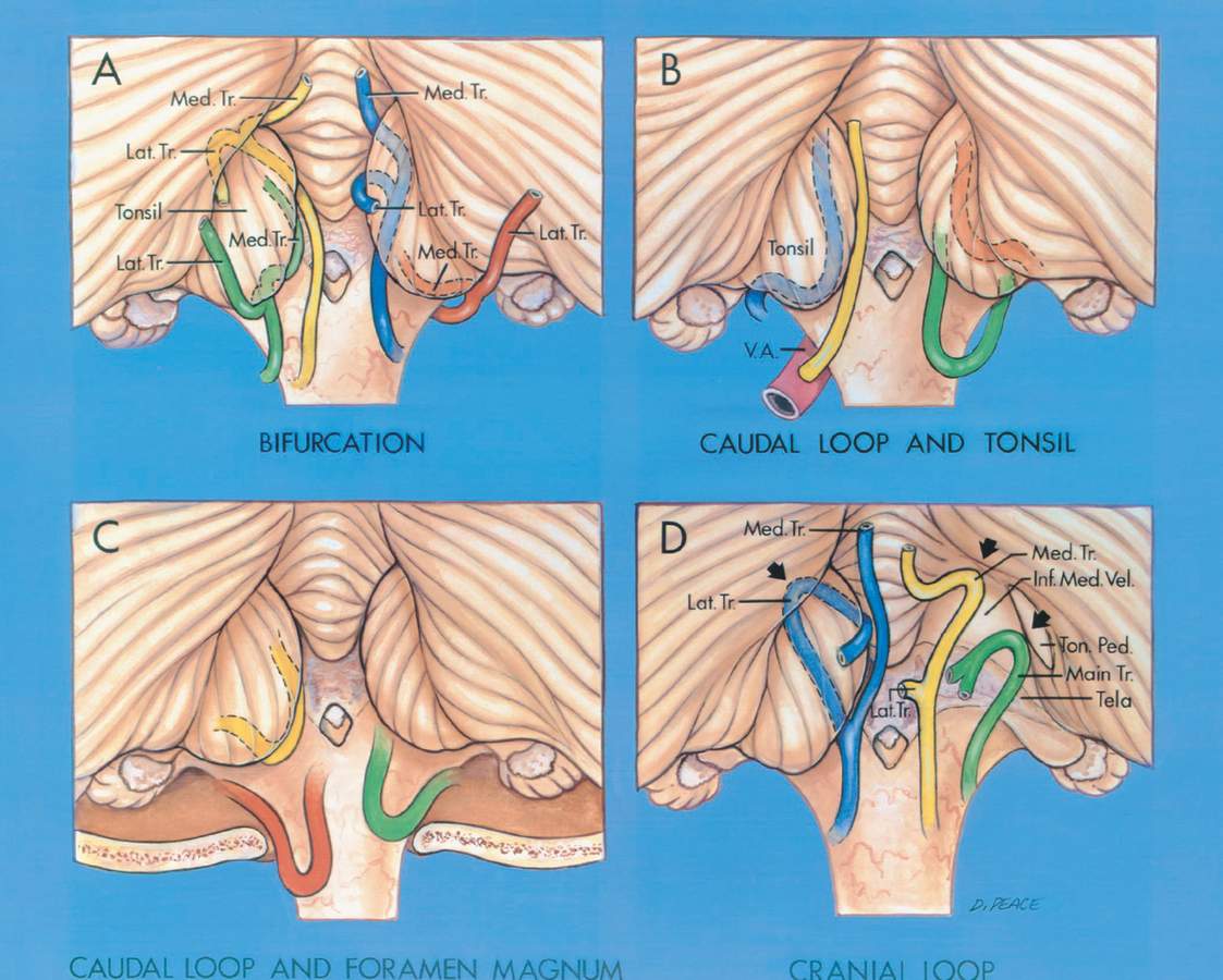
Locations of the Posterior Inferior Cerebellar Artery Bifurcation, the Caudal Loops, and the Cranial Loops
5637
Surgical Correlation
Tags
A, Site of the bifurcation in relation to the tonsil. The main trunk of the PICA may bifurcate at any site along the margin of the tonsil. Inferolateral bifurcation (red): the lateral trunk passes upward lateral to the tonsil to reach the hemisphere, and the medial trunk passes along the anteromedial margin of the tonsil. Inferomedial bifurcation (green): the lateral trunk passes superolateral over the posterior margin of the tonsil to reach the hemispheric surface, and the medial trunk passes upward along the anteromedial margin of the tonsil. Superomedial bifurcation (blue): the lateral trunk passes posteriorly over the medial surface of the tonsil, and the medial trunk ascends to supply the vermis. Superolateral bifurcation (yellow): the lateral trunk passes out of the fissure between the tonsil and the hemisphere and proceeds to the hemispheric surface, and the medial trunk ascends to supply the vermis. B, Location of the caudal loop in relation to the tonsil. The tonsillomedullary segment often formed a caudally convex loop (blue, orange, green) as it passed medially across the posterior surface of the medulla. This caudal part of the tonsillomedullary segment was located between 10.0 mm inferior and 13.0 mm superior (average, 1.6 mm superior) to the caudal tip of the tonsil. This loop could be found superior to (orange), inferior to (green), or at the level of (blue) the caudal tip of the tonsil. In some cases (yellow), the PICA ascended from the vertebral artery (V.A.) or took another course to reach the medial surface of the tonsil without forming a caudal loop. C, Relation of the caudal loop to the foramen magnum. Most caudal loops were superior to the foramen magnum (yellow), but they could be inferior to (red) or at the level of (green) the foramen magnum. The caudal loop was located between 7.0 mm inferior and 18.0 mm superior (average, 6.9 mm superior) to the foramen magnum. D, Relationship of the cranial loop (arrow) to the superior pole of the tonsil and the trunks of the PICA. The right tonsil was removed at the level of the tonsillar peduncle to expose the inferior medullary velum and the tela choroidea. The telovelotonsillar segment often formed a cranially convex loop. below the fastigium. The cranially convex loop could be formed by either the main (green), medial (yellow), or lateral (blue) trunk. On the left (blue), the lateral trunk (arrow) forms a cranially convex loop over the superior pole of the tonsil and the medial trunk ascends straight to the vermis. In the center (yellow), the medial trunk (arrow) forms a cranially convex loop at the superior pole of the tonsil and the lateral trunk courses around the medial surface of the tonsil. On the right (green), the cranial loop is formed by the main trunk (arrow) and lies in the telovelotonsillar fissure anterior to the superior pole of the tonsil. (Images courtesy of AL Rhoton, Jr.)


