

Developmental Venous Anomaly

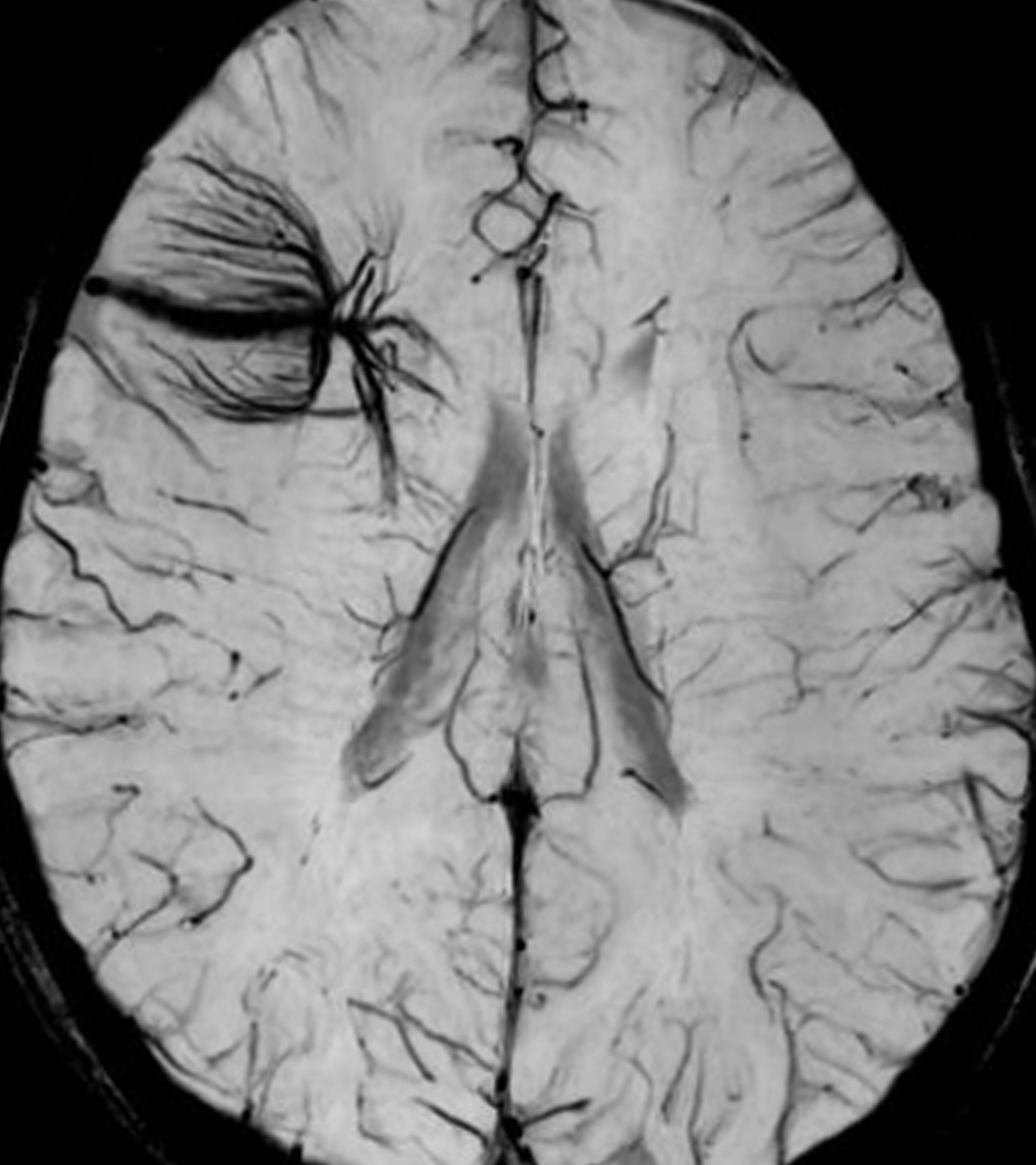
Figure 1: Axial susceptibility-weighted imaging (SWI) reveals a cluster of hypointense vessels emerging from the right frontal lobe cortex and deep white matter and draining into an anomalous, asymmetrically enlarged cortical vein.
Overview
Developmental venous anomalies (DVAs), also known as venous angiomas in the literature, are mature venous structures with abnormal (anomalous) but nonpathologic drainage patterns. These frequently coexist with cavernous malformations (CMs), capillary malformations, and venolymphatic malformations but are usually not anatomically related. DVAs have been implicated in epilepsy, sometimes in association with areas of cortical dysplasia.
Imaging
- Umbrella-like configuration of enlarged medullary veins converging (draining) into an enlarged vein, either into dural sinuses or deep ependymal veins
- Often emanating from a ventricle
- Typically solitary and of variable length (<2–3 cm)
- Clinically silent in vast majority of cases
- Hemorrhage is rare, but risk increases if associated with malformations or if draining vein is thrombosed
- Associations
- Up to 20% coexist with CM or capillary telangiectasias
- Blue rubber bleb nevus syndrome
- Venolymphatic malformation
- Proposed association with sulcation-gyration abnormalities and epilepsy
- Nonenhanced CT
- Typically not visible
- Occasionally calcified if mixed or associated directly with CM
- Rarely, acute parenchymal hemorrhage (if draining vein spontaneously thromboses)
- CT angiography
- Numerous linear or dot-like enhancing foci
- Well-circumscribed, round/ovoid, enhancing areas on sequential sections
- Converge on single enlarged tubular draining vein
- Occasionally seen as linear structure in single slice
- MRI
- Multiple pulse sequences available to evaluate various tissue properties as well as dynamic properties (flow, velocity)
- MRI accurately defines the vascular channels forming DVAs, having the ability to detect vascular flow
- Generally does not require intravenous contrast to detect
- Findings
- T1WI
- Can be normal if DVA is small
- Variable signal depending on size; a small flow void can often be detected
- T2WI
- Black flow voids
- Linear, umbrella-like configuration of dark small vascular flow voids
- FLAIR
- Often normal
- Hyperintense vessel may be visible
- May show hyperintense region if venous ischemia or hemorrhage present
- Possible to have colateralization with electroencephalographic findings in epilepsy evaluation
- T2* GRE and SWI
- Hypointense blooming artifact on GRE if large or if coexisting CM with hemorrhage
- Usually clearly visible hypointense vessels on SWI due to blood oxygen level-dependent (BOLD) effect in draining veins
- DWI
- Usually normal
- T1WI C+
- Strong enhancement of numerous linear or dot-like foci converge on single, asymmetrically enlarged vein, draining into dural sinuses or deep ependymal veins
- T1WI
- MRA
- Findings
- Arterial phase usually normal
- Contrast-enhanced MRA may demonstrate slow-flow DVA
- MR venography may delineate umbrella configuration and drainage pattern
- No indication for catheter angiography
- Findings
- Digital subtraction angiography
- DVA is an incidental finding in the vast majority of cases at cross-sectional imaging and is not an indication for catheter angiography
- Arterial phase normal in >95% of cases
- Venous phase: radially oriented, dilated medullary veins in an umbrella-like configuration, sometimes emanating from the ventricle

Figure 2: Six-year-old girl with seizure—“evaluate for AVM.” (Left) An MRI series identifies an abnormal cluster of vessels emerging from the left subinsular and left lentiform nuclei and converging at or the near the ependymal surface of the left lateral ventricle. (Right) Oblique projection DSA image. Left internal carotid injection (venous phase image) confirms a developmental venous anomaly with umbrella-like configuration of anomalous subinsular veins coalescing into a prominent subependymal vein without early venous opacification to suggest high flow shunt.
For more information, please see the corresponding chapter in Radiopaedia.
Contributor: Daniel Murph, MD
References
Oran I, Kiroglu Y, Yurt A, et al. Developmental venous anomaly (DVA) with arterial component: a rare cause of intracranial haemorrhage. Neuroradiology 2009;51:25–32. doi.org/10.1007/s00234-008-0456-9
Abla A, Wait SD, Uschold T, et al. Developmental venous anomaly, cavernous malformation, and capillary telangiectasia: spectrum of a single disease. Acta Neurochir (Wien) 2008;150:487–489. doi.org/10.1007/s00701-008-1570-5
Fushimi Y, Miki Y, Togashi K, et al. A developmental venous anomaly presenting atypical findings on susceptibility-weighted imaging. AJNR Am J Neuroradiol 2008;29:E56. doi.org/10.3174/ajnr.A1074
Pozzati E, Marliani AF, Zucchelli M, et al. The neurovascular triad: mixed cavernous, capillary, and venous malformations of the brainstem. J Neurosurg 2007;107:1113–1119. doi.org/10.3171/JNS-07/12/1113
Please login to post a comment.


