The Basal Vein and Its Tributaries
6850
Surgical Correlation
Tags
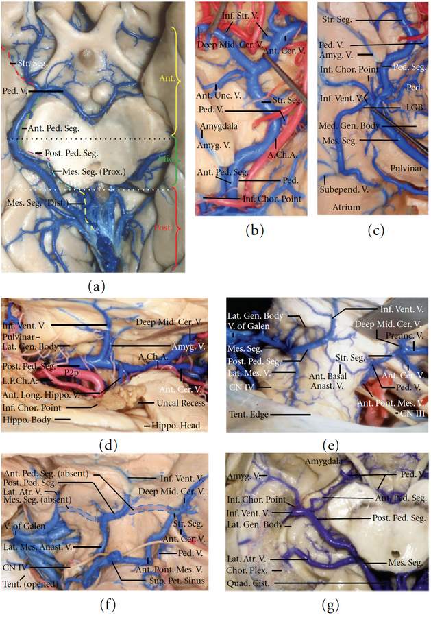
The Basal Vein and Its Tributaries. a, Inferior view. The basal vein and its tributaries have been exposed. The segmental classification of the basal vein (right hemisphere) and the segmental classification of the MTR (left hemisphere) are shown. The striate (red interrupted line) and anterior peduncular (green interrupted line) segments of the basal vein are located along the anterior MTR (yellow bracket). The posterior peduncular (purple interrupted line) and proximal mesencephalic (aqua interrupted line) segments of the basal vein are situated in the middle MTR (green bracket). The distal mesencephalic segment (yellow interrupted line) of the basal vein belongs to the posterior MTR (red bracket). The anterior peduncular segment is also referred to as the anterior anastomotic vein, because it allows the striate segment to communicate with the posterior peduncular segment. The mesencephalic segment of the basal vein also is referred to as the posterior basal anastomotic vein because it connects the peduncular segment to the vein of Galen. b, Basal view of the striate and anterior peduncular segments of the basal vein. The part of the amygdala below the level of the optic tract has been removed. The carotid and crural cisterns have been opened to expose the striate and anterior peduncular segments of the basal vein. The anterior and deep medial cerebral veins meet to form the striate segment of the basal vein. The anterior peduncular segment begins where the peduncular vein joins the basal vein, and the posterior peduncular segment starts at the junction with the inferior ventricular vein. c, Inferior view of the roof of the temporal horn in another right hemisphere. The junctional point between the inferior ventricular vein and the peduncular segment corresponds to the cisternal level of the inferior choroidal point and the junction of the crural and ambient cisterns. Several large subependymal veins draining the roof and lateral wall of the temporal horn join to form the inferior ventricular vein. d, The right temporal horn has been opened, and the ambient and crural cistern have been exposed by opening the choroidal fissure. The amygdalar vein drains separately from the inferior ventricular vein into the basal vein. The anterior longitudinal hippocampal vein empties into the basal vein anterior to the inferior ventricular vein. e, Lateral view of the right tentorial incisura. The temporal lobe has been removed to expose the basal vein. The anterior peduncular segment of the basal vein is hypoplastic. Failure of anastomosis between the striate and peduncular segments of the basal vein results in formation of a prominent preuncal vein that drains forward into the sphenoparietal sinus. f, Same view as e in a different specimen. Both the anterior peduncular (anterior anastomotic vein) and the mesencephalic segment (posterior anastomotic veins; blue shaded areas) are absent, and the striate, peduncular, and mesencephalic segments of the basal vein are disconnected from each other. The striate segment drains inferiorly via the anterior pontomesencephalic vein, the peduncular segment drains inferiorly through a prominent lateral mesencephalic vein, and the tributaries of the mesencephalic segment drain directly into the vein of Galen. g, Inferior view of the right basal vein. The anterior peduncular segment (anterior anastomotic vein) of the basal vein is hypoplastic. The inferior ventricular vein empties into and forms the posterior peduncular and mesencephalic segments of the basal vein, and the anterior segment of the basal vein drains anteriorly. A prominent lateral atrial vein drains into the mesencephalic segment. (Images courtesy of AL Rhoton, Jr.)


