The Atlas
5945
Surgical Correlation
Tags
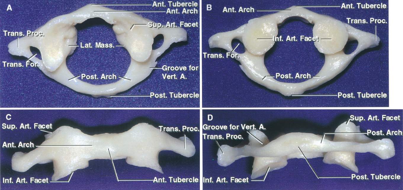
The atlas consists of two thick lateral masses situated at the anteromedial part of the ring, which are connected in front by a short anterior arch and posteriorly by a longer curved posterior arch. The anterior and posterior tubercles are at the anterior and posterior mid-line. The superior articular facet is an oval, concave facet that faces upward and medially to articulate with the occipital condyle. The inferior articular facet is a circular, flat, or slightly concave facet that faces downward, medially, and slightly backward and articulates with the superior articular facet of the axis. The medial aspect of each lateral mass has a small tubercle for the attachment of the transverse ligament of the atlas. The transverse process projects from the lateral masses. The transverse foramina transmit the vertebral arteries. The upper surface of the posterior arch adjacent to the lateral masses has paired grooves in which the vertebral arteries course. A, Superior view. B, Inferior view. C, Anterior view. D, Posterior view. (Images courtesy of AL Rhoton, Jr.)


