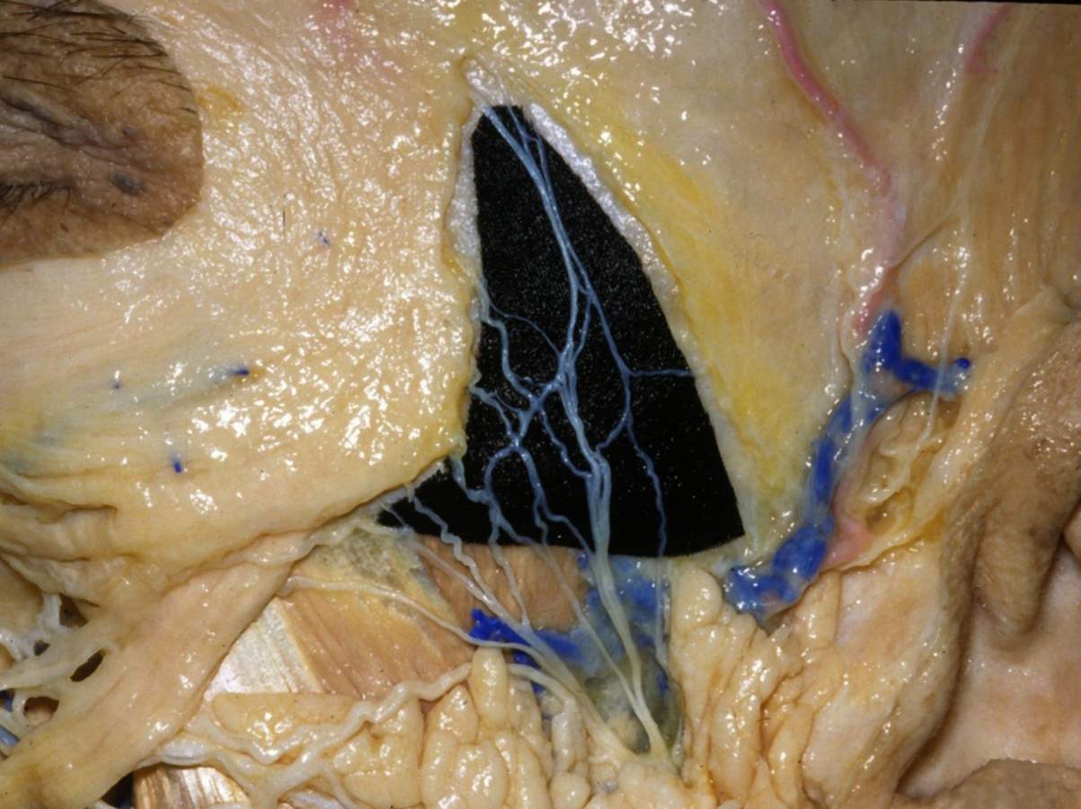
Temporoparietal Fascia Covering Surface of Left Side of Head
7221

Surgical Correlation
Tags
Temporoparietal fascia covering surface of left side of head. The temporoparietal fascia lies deep to the dermis of the skin and covers the temporalis muscle and its deep fascia. The temporoparietal fascia is continuous below the zygomatic arch with the superficial musculoaponeurotic system of the face and with the galea aponeurotica covering the vertex of the skull. It receives from below the superficial temporal artery and vein and auriculotemporal nerve (seen in front of the ear where they cross the root of the zygomatic arch). A triangular piece of black fabric has been placed beneath the frontal or temporal branches and some of the zygomatic branches of the facial nerve. These emerge from the border of the parotid gland and course on the undersurface of the temporoparietal fascia to the frontalis and orbicularis oculi muscles, respectively. Buccal branches of the facial nerve cross the masseter muscle parallel with the parotid (Stensen's) duct to facial muscles of the central face. (Image courtesy of AL Rhoton, Jr.)


