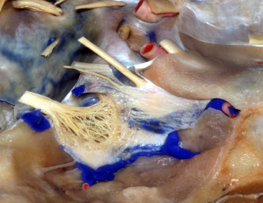
Superolateral View of Right Cavernous Sinus Wall
6306

Surgical Correlation
Tags
Superolateral view of right cavernous sinus wall. The meningeal dura has been removed over the lateral wall of the cavernous sinus to reveal several cranial nerves that course to the orbit. Entering the edge of the tentorium cerebelli attachment to the anterior and posterior clinoid processes are the oculomotor and trochlear nerves (the former located superior to the trochlear nerve). Inferior to the trochlear nerve is the ophthalmic nerve. All three of these nerves enter the orbit through the superior orbital fissure, a space between the lesser and greater wings of the sphenoid. Along the posteroinferior border of the lesser wing is the sphenoparietal sinus, a small sinus, that drains into the cavernous sinus. The trigeminal ganglion occupies Meckel's cave. The maxillary nerve is shown entering the foramen rotundum and the mandibular nerve the foramen ovale. Posterolateral to this foramen is foramen spinosum transmitting the middle meningeal artery. The greater superficial petrosal nerve is shown leaving its hiatus on the anterior surface of the petrous bone and passing deep to the trigeminal ganglion. (Image courtesy of AL Rhoton, Jr.)


