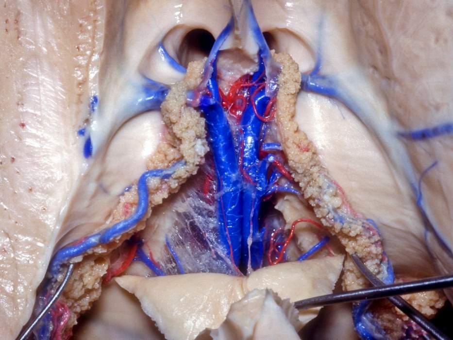
Superior View of Velum Interpositum
5436

5436

Superior view of the lateral and third ventricles through the velum interpositum after partial resection of the fornices. The choroid plexus of both lateral ventricles is visible entering the foramina of Monro. At this level, the paired septal veins merge with the thalamostriate veins to form both internal cerebral veins, which course posteriorly along the roof of the third ventricle. Choroidal veins and arteries are visible. (Image courtesy of AL Rhoton, Jr.)
Check to see if you have access through your library or institution.
Start your 30-day free trial or subscribe to access the most comprehensive collection of advanced microneurosurgical techniques. The Neurosurgical Atlas collection presents the nuances of technique for complex cranial and spinal cord operations.
Login or registerCopyright © 2024 Neurosurgical Atlas, Inc. All Rights Reserved. | End User License Agreement | Privacy Policy | Report a Problem
