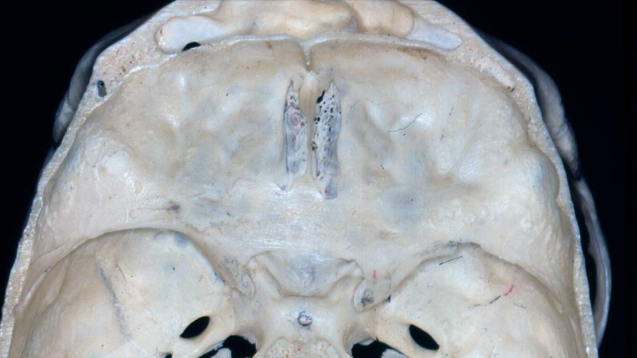
Superior View of the Bony Anterior and Middle Cranial Fossae
7540

Surgical Correlation
Tags
Superior view of the bony anterior and middle cranial fossae. The anterior cranial fossa forms the anterior floor of the neurocranium and consists of the frontal bone, cribriform plate and crista galli of the ethmoid bone, planum sphenoidale, and lesser wings of the sphenoid. The cribriform plate is sieve-like for transmission of neurofilaments of the olfactory nerves to the olfactory bulbs. The frontal sinus is a paranasal space between the outer and inner tables of compact frontal bone anteriorly. The medial portions of the lesser wings end as curved projections, the anterior clinoid processes, medial to which are the openings of the optic canals. Posterior to the anterior fossa is the middle fossa, a depression bounded anteriorly by the lesser sphenoidal wings and posteriorly by the petrous portion of the temporal bone. The body of the sphenoid is located in the midline. The floor of the middle fossa is formed by the greater wings of the sphenoid and the squamous portion of the temporal bone. A crescent of foramina are related to the greater wings, including the superior orbital fissure, foramen rotundum, foramen ovale, a foramen spinosum. The foramen lacerum is a space located at the junction of the sphenoid body, greater wing, and apex of the petrous bone. Its floor is filled by fibrocartilage in life. The body of the sphenoid superiorly features a pair of transverse ridges that border a depression, the hypophyseal fossa, for the pituitary gland. The anterior ridge is the tuberculum sellae, the posterior ridge is the dorsum sellae. The body of the sphenoid contains the sphenoid sinuses. (Image courtesy of PA Rubino)


