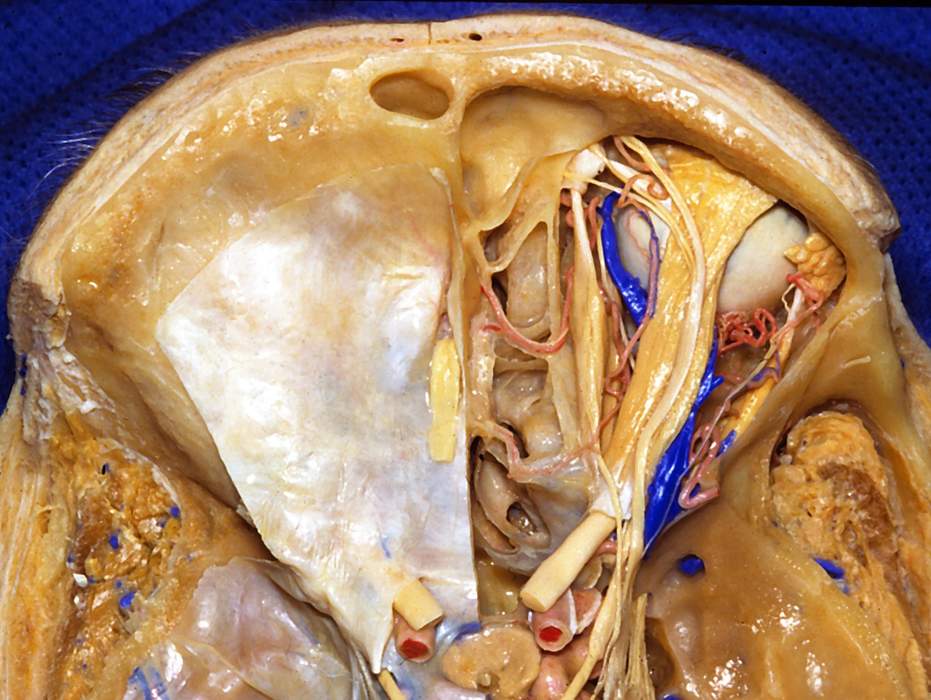
Superior View of Contents of Right Orbit
6507

Surgical Correlation
Tags
Superior view of contents of right orbit. The orbital plate of the frontal bone and periorbita have been removed to expose contents of the orbit and bone has been removed to expose the frontal, ethmoid, and sphenoid sinuses as well. The eyeball or globe occupies the anterior half of the orbit while the extraocular muscles, fat, and neurovascular elements populate the posterior half. The latter structures enter the apex of the orbit through the optic canal (for the optic nerve and ophthalmic artery) and the superior orbital fissure (for the oculomotor, trochlear, ophthalmic, and abducens nerves, and the superior ophthalmic vein). A superficial strata of extraocular muscles can be seen: superior oblique muscle follows the medial wall, lateral rectus muscle follows the lateral wall, and the levator palpebrae superioris muscle courses forward between these to the upper eyelid. The superior rectus muscle lies deep to the levator, and the medial rectus muscle lies deep to the superior oblique. The recti muscles attach in common to the annulus of Zinn or tendinous ring at the orbital apex. The optic, oculomotor (both superior and inferior divisions), abducens, and nasociliary nerves and the ophthalmic artery pass through this ring to enter the orbit while the frontal, lacrimal, and trochlear nerves and the superior ophthalmic vein do not. The ophthalmic artery, a branch of the internal carotid artery, enters the orbit with the optic nerve and gives rise to the lacrimal artery before crossing the optic nerve, in company with the nasociliary nerve, toward the medial side of the orbit. Here, it gives rise to the supraorbital, and posterior and anterior ethmoidal branches. The latter pass through foramina on the medial wall of the orbit to supply ethmoid and sphenoid sinuses before entering the nasal cavity. The ophthalmic nerve parallels the early course of the maxillary nerve (which passes to the foramen rotundum) before dividing into its "NFL" branches (nasociliary, frontal, lacrimal), which then enter the orbit through the superior orbital fissure. The lacrimal nerve courses along the lateral wall of the orbit (near the superior border of the lateral rectus) to the lacrimal gland. The frontal nerve courses forward in the orbit superficial to the levator palpebrae superioris and superior rectus muscles before dividing into the supraorbital (the larger of the two) and supratrochlear branches. At the apex of the orbit, the trochlear nerve bends medially (medial to the frontal nerve) to enter the superior border of the superior oblique muscle. The superior ophthalmic vein provides the principal venous drainage from the orbit and empties into the cavernous sinus. The vein is larger in diameter than the ophthalmic artery and the artery presents a greater tortuosity in appearance. (Image courtesy of AL Rhoton, Jr.)


