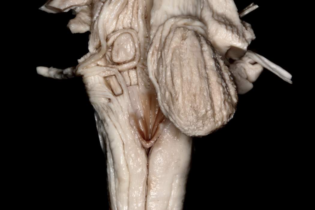
Superficial White Matter Dissection of the Fourth Ventricular Floor
6529

Surgical Correlation
Tags
Superficial white matter dissection of the fourth ventricular floor. The stria medullaris (not seen in this dissection) divides the rhomboid fossa (floor of the fourth ventricle) into two triangular areas: a superior or pontine, and an inferior or medullary (open medulla oblongata). Inferiorly, the fourth ventricle narrows at the obex and continues caudally as the central canal. The obex is located in the posterior midline between the cranial ends of the Gracilis tubercles. On the medullary area of the fourth ventricle floor, there are two noticeable trigones: the hypoglossal (more cranial and medial) formed by the hypoglossal nucleus, which contains general somatic efferent neurons; and the vagal (more lateral) formed by the dorsal motor nucleus of the vagus, which contains visceral parasympathetic neurons. The apices of both trigones point caudally, forming the calamus scriptorium as it is shaped as a writing quill nib. The facial colliculus is a very important prominence on the pontine triangle and is formed by the first genu of the facial nerve coursing behind the motor nucleus of the abducens nerve. (Image courtesy of AL Rhoton, Jr.)


