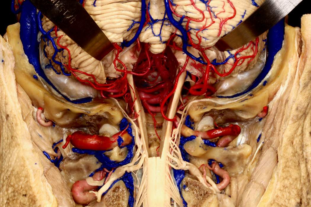
Suboccipital Craniectomy with Cerebellum Retracted
6526

Surgical Correlation
Tags
Suboccipital craniectomy with cerebellum retracted. In this image, the occiput and associated musculoskeletal structures have been removed to visualize a wide suboccipital craniectomy. The cerebellum is retracted superiorly, allowing visualization of the upper cervical spinal cord, the medulla oblongata and the pons (which have been hollowed to reveal a window through to the vertebrobasilar junction). The vertebral arteries are visible coursing medially along the superior edge of the C1 vertebra after passing though the foramen tranversarium. The spinal accessory nerve fibers ascend from the cervical spinal cord intracranially to exit the skull base via the jugular foramen with the glossopharyngeal and vagus nerves and the sigmoid sinus. Prominent C2 nerve roots are apparent here with large dorsal root ganglia, supplying occipital nerves. (Image courtesy of AL Rhoton, Jr.)


