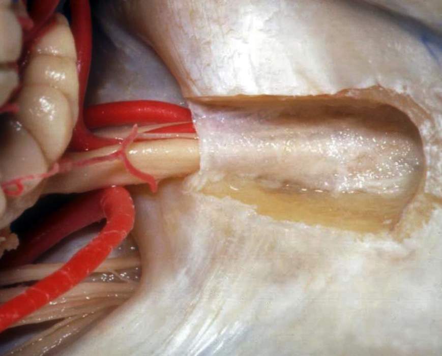
Right Posterosuperior Views of the Internal Auditory Canal and Jugular Foramen
6522

Surgical Correlation
Tags
Right posterosuperior views of the internal auditory canal and jugular foramen. From beneath the lateral margin of the cerebellum appear the anterior inferior cerebellar artery and the facial and vestibulocochlear nerves. The latter course toward the opening (porous) of the internal auditory meatus. A portion of the roof is its canal has been drilled. The nerves are accompanied by the labyrinthine artery, a branch of the anterior inferior cerebellar artery. It supplies the entire inner ear. The subarcuate artery, usually a branch of the labyrinthine or anterior inferior cerebellar artery, courses toward the subarcuate fossa, a tiny opening superolateral to the internal auditory canal. Here, it enters the petrous bone supplying it and the bony labyrinth. A portion of the posterior inferior cerebellar artery can be seen at the lower left border of this image in relation to the glossopharyngeal, vagus, and spinal accessory nerves. These nerves can be seen entering the jugular foramen. (Image courtesy of AL Rhoton, Jr.)


