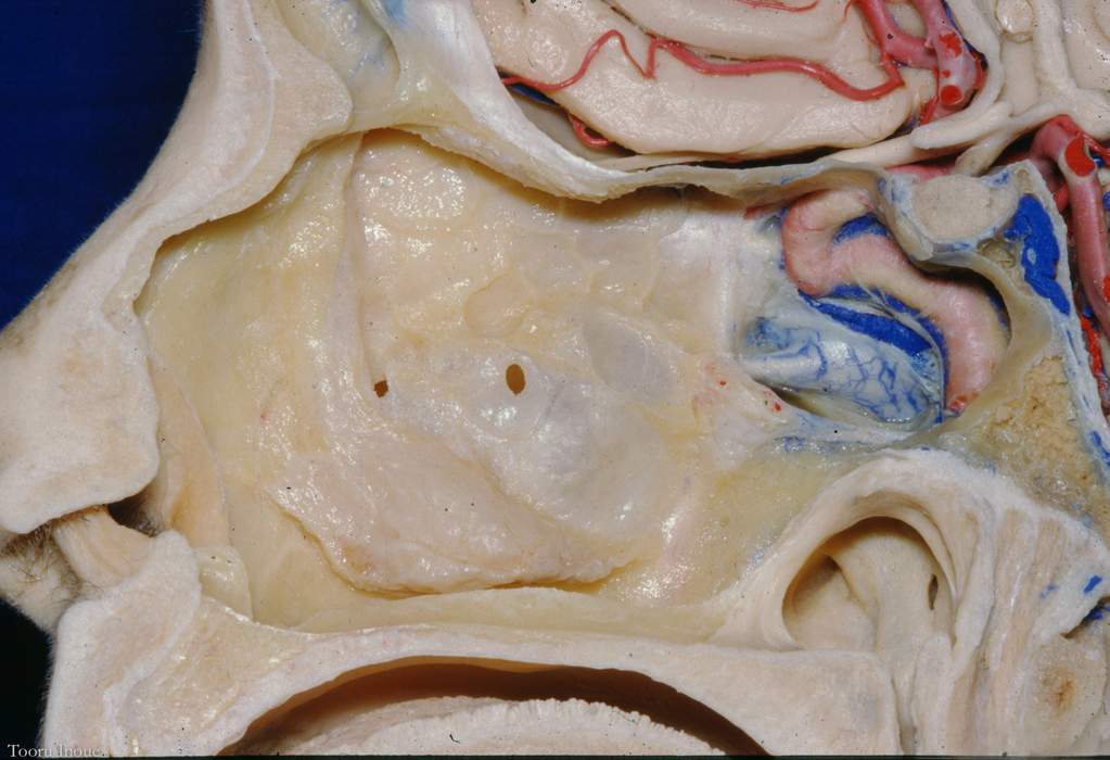
Right Lateral Wall of the Nasal Cavity, Nasopharynx, and Cavernous Sinus
5806

Surgical Correlation
Tags
Right lateral wall of the nasal cavity, nasopharynx, and cavernous sinus. The superior and middle turbinates, components of the ethmoid bone, have been removed from the lateral wall of the nasal cavity. The inferior turbinate, a separate bone, remains intact. The nasal cavity is separated from the oral cavity by the hard palate, consisting of the palatine processes of the maxilla and horizontal plates of the palatine bones. Posterior to the inferior turbinate is the torus tubarius projecting into the nasopharynx. Projecting inferiorly from the torus is the salpingopharyngeal fold containing the salpingopharyngeus muscle. Posterior to this fold is the pharyngeal recess, which is bounded posteriorly by the superior pharyngeal constrictor. Emerging from the floor of the opening of the Eustachian tube is the salpingopalatine fold or torus levatorius. Deep to its mucosal lining is the levator veli palatini muscle. The body of the sphenoid has been removed to visualize the cavernous sinus surrounding the cavernous segment of the internal carotid artery and its carotid nerve plexus. The pituitary gland is shown resting in the hypophyseal fossa and the frontal lobes of the cerebrum and associated vasculature occupy the anterior cranial fossa. (Image courtesy of AL Rhoton, Jr.)


