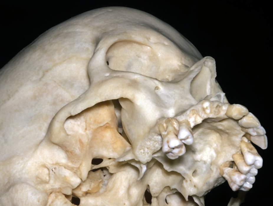
Right Anterolateral Inferior View of Skull
6517

Surgical Correlation
Tags
Right anterolateral inferior view of skull. The mandible has been removed to allow view of the temporal and infratemporal fossae. The head or condyle of the mandible resides in the mandibular fossa. This fossa is bounded anteriorly by the articular tubercle at the anterior root of the zygomatic process of the temporal bone and posteriorly by the tympanic part of the temporal bone. The latter contains the external auditory canal or meatus. The squamous portion of the temporal bone is a "scale-like" piece of bone that forms much of the temporal fossa and includes the zygomatic process. This process unites with the temporal process of the zygomatic bone to form the zygomatic arch. Superior to the squamous temporal bone is the parietal bone. They unite at the squamosal or temporoparietal suture. Anterior to the parietal bone is the frontal bone, which join at the coronal suture. Inferior to the lateral aspect of the frontal bone is the greater wing of the sphenoid bone. The junction of these four bones is the pterion, which marks the location of the anterior branch of the middle meningeal artery internally. The zygomatic bone forms the prominence of the cheek. The horizontal plane through the zygomatic arch marks the boundary between the temporal and infratemporal fossae. This latter space is bounded medially by the lateral plate of the pterygoid process of the sphenoid bone. Its roof is formed by the infratemporal surface of the greater wing of the sphenoid and contains the foramen ovale and foramen spinosum. The infratemporal fossa communicates with the pterygopalatine fossa medially through the pterygomaxillary fissure, a vertical space between the pterygoid process and posterior wall of the maxilla. Both fossae communicate with the floor of the orbit via the inferior orbital fissure, a space between the maxillary bone and greater wing of the sphenoid. The maxilla is the bone of the central part of the face. It forms the floor and inferior margin of the orbit, bounds much of the anterior nasal or piriform aperture of the nasal cavity, contains the maxillary teeth, and forms the anterior two-thirds of the hard palate. The horizontal part of the palatine bone forms the posterior one-third of the palate. The infraorbital foramen is an opening in the maxilla a few millimeters inferior to the lower orbital rim for transmission of the infraorbital neurovasculature to the lower eyelid, lateral nose, and upper lip. The nasal bones are narrow paired bones that articulate with the frontal bone superiorly and with the maxillary bones lateroinferiorly. They form the superior boundary of the piriform aperture of the nasal cavity. (Image courtesy of AL Rhoton, Jr.)


