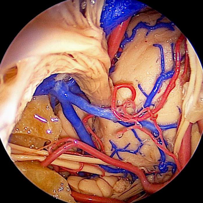
Magnified Endoscopic View of Right Anterior Aspect of Petroclival Area
6354

Surgical Correlation
Tags
Magnified endoscopic view of right anterior aspect of petroclival area. Petrosal and clival bone have been removed to expose the anterolateral pons. The abducens nerve is shown ascending from the lower ventral pons through a remnant of clival dura. The anterior inferior cerebellar artery can be seen arising from the proximal portion of the basilar artery and looping toward the internal auditory meatus in company with the facial and vestibulocochlear nerves. The trigeminal nerve emerges from the lateral pons and courses toward the petrous apex. Posterior and inferior to it is the superior petrosal vein, an important venous channel draining the anterior cerebellum and brainstem. It is formed by union of several veins, including the transverse pontine vein, vein of the middle cerebellar peduncle, vein of the cerebellopontine fissure, the pontine trigeminal vein, and anterior lateral marginal vein. The superior petrosal vein empties into the superior petrosal sinus. (Image courtesy of AL Rhoton, Jr.)


