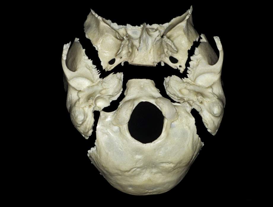
Inferior Perspective of Disarticulated Skull Base
5550

Surgical Correlation
Tags
Inferior perspective of disarticulated skull base. This image presents the inferior views of the unpaired sphenoid and occipital bones and the paired temporal bones. The occipital bone forms the posterior base of the skull while the temporal bones and sphenoid form the middle region. The foramen magnum in the occipital bone is the largest foramen in the skull for passage of the spinal cord and dura into the vertebral canal, the vertebral arteries, and spinal accessory nerves. The jugular foramen is an opening between the occipital bone and petrous portion of temporal that allows the internal jugular vein and cranial nerves IX, X, and XI to exit the skull. Foramen lacerum is an opening between the apex of the petrous bone, body of sphenoid, and the basilar portion of the occipital. It is filled with fibrocartilage in life. The petroclival fissure is a groove between the petrous bone and clivus (a slope of bone anterior to the foramen magnum formed by union of the dorsum sellae and basilar portion of the occipital) which contains the inferior petrosal sinus that drains the cavernous sinus to the internal jugular vein through the jugular foramen. (Image courtesy of AL Rhoton, Jr.)


