Frontalis Fascial Planes and Dissection
6977
Surgical Correlation
Tags
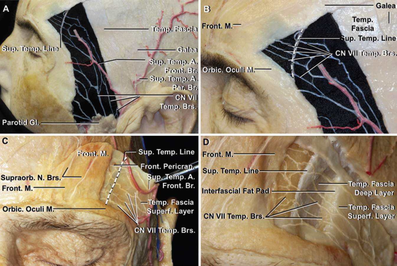
Frontalis Fascial Planes and Dissection. A, The skin, subcutaneous tissues, and galea have been removed to expose the facial branches coursing in the loose areolar tissue on the outer surface of the temporal fascia. The trunks of the facial nerve exiting the upper edge of the parotid gland and crossing the outer surface of the temporal fascia and frontal pericranium are exposed. Black material has been placed deep to the small branches of CN VII to highlight their course in the loose areolar tissue on the outer surface of the temporal fascia. There are commonly 3 temporal trunks—anterior, middle, and posterior—at the level of the zygomatic arch. The anterior and middle temporal branches supply the orbicularis and frontalis muscles. The posterior temporal branch commonly supplies the auricularis muscle. B, Enlarged view showing the 3 temporal branches at the level of the parotid gland have divided into 5 tiny branches as they cross the STL (interrupted line). C and D, Stepwise dissection shows the relationship of the nerves to the frontalis muscle to the superficial layer of temporal fascia, frontal pericranium, lateral edge of the frontalis muscle, and STL. C, Anterior view. The skin and subcutaneous tissues and galea have been removed to expose the nerves to the frontalis muscle coursing in the loose areolar layer on the outer surface of the superficial layer of temporal fascia and frontal pericranium. The nerves supplying the frontalis muscle pass below the lateral edge of the anterior part of the muscle. The superficial layer of temporal fascia attaches at the STL (interrupted line) and blends medially into the frontal pericranium. The supraorbital branches of CN V ascend on the outer surface of the frontalis muscle. D, A window in the fascial layers adjoining the STL has been completed by removing the superficial layer of temporal fascia and the frontal pericranium to show the nerves to the frontalis muscle crossing the STL. The temporal fascia at and below the level of the upper edge of the interfascial fat pad splits into deep and superficial layers that enclose it. (Images courtesy of AL Rhoton, Jr.)


