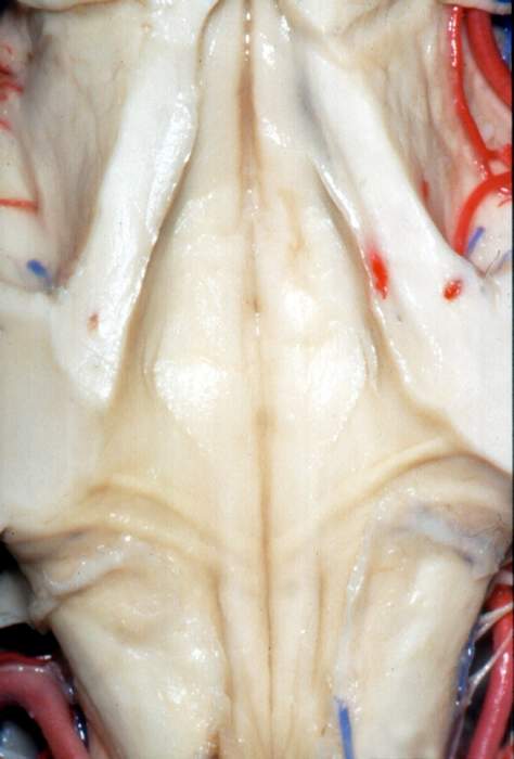
Floor of the Fourth Ventricle
6438

Surgical Correlation
Tags
Floor of the fourth ventricle. Superior view of the fourth ventricle. The cerebellum, superior and inferior medullary velums have been removed to reveal the floor of the fourth ventricle. The middle and superior cerebellar peduncles has been sectioned in a coronal plane. In the floor of the fourth ventricle at the level of the caudal pons are the paired facial colliculi, formed by facial nerve fibers wrapping around the abducens nucleus. In the floor of the ventricle at the level of the medulla lie the slender, elongated hypoglossal and vagal trigones, formed by the underlying hypoglossal nucleus and dorsal motor nucleus of the vagus, respectively. Just behind the middle cerebellar peduncle, the ventricle tapers on either side to form the lateral recesses, which become continuous with the lateral apertures, or Foramina of Luschke. (Image courtesy of AL Rhoton, Jr.)


