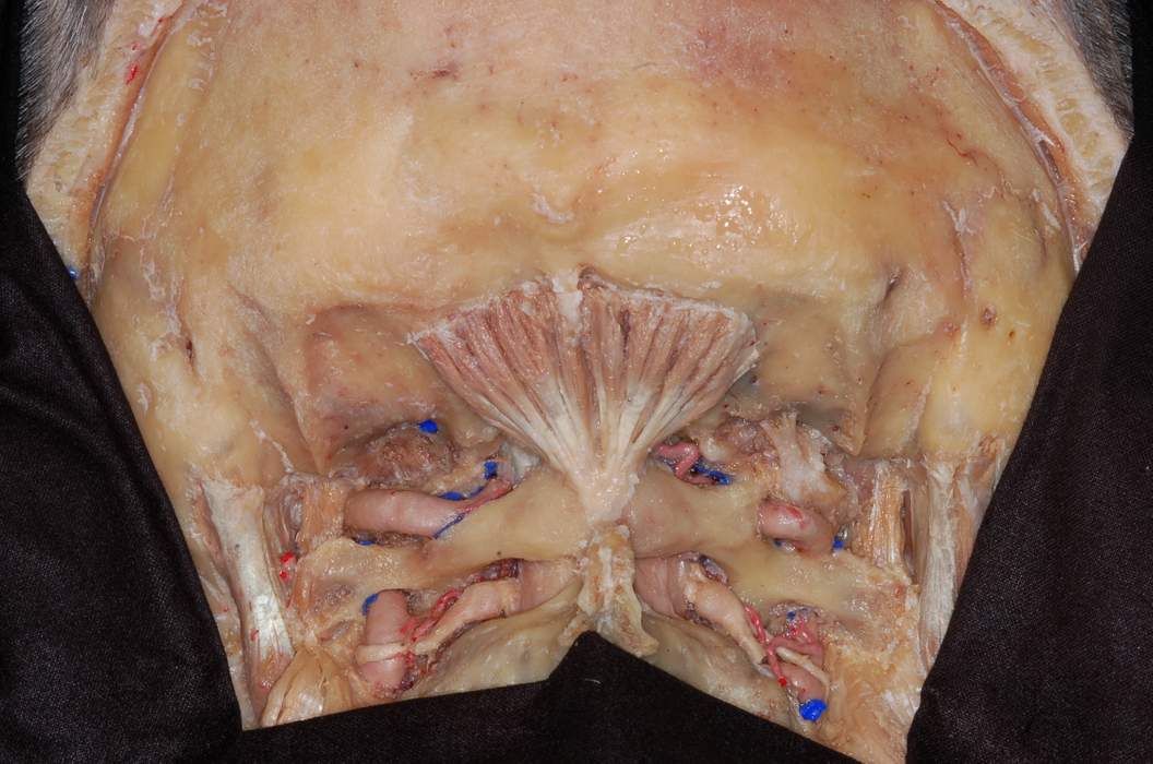
Atlantic Part of the Vertebral Artery
7460

Surgical Correlation
Tags
Atlantic Part of the Vertebral Artery. The atlantic part of the vertebral artery is shown emerging from the transverse foramen of C2, coursing upward through the transverse foramen of C1, and around the lateral mass to occupy a groove on the superior surface of the posterior arch of the atlas. The rectus capitis posterior minor muscles arise from the posterior tubercle and attach to the medial half of the inferior nuchal line. Inferior to the posterior arch is the lamina of the axis. The elastic ligamenta flava bridge the gap between these bony parts contributing to the roof of the vertebral canal. Emerging between the C1 and C2 vertebrae are the C2 spinal nerves. These are formed by union of ventral and dorsal roots. The distal part of the latter shows a swelling, the dorsal root or spinal ganglion, containing the cell bodies of sensory neurons associated with this nerve. The definitive spinal nerve is short and exits the intervertebral foramen where it subsequently divides into ventral and dorsal primary rami. (Image courtesy of PA Rubino)


