Approach to the Upper Clivus
6704
Surgical Correlation
Tags
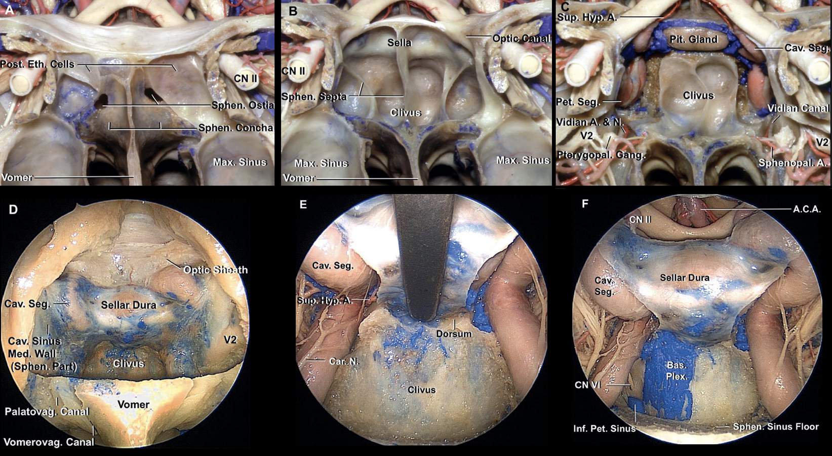
Approach to the Upper Clivus. A through C, Relationships of the sphenoid sinus and clivus. A, Anterior view of the sphenoid sinus. The posterior ethmoid air cells have a common wall with the superolateral face of the sphenoid sinus and extend along the lateral edge of the sphenoid ostia. The portion of the sphenoid face below and between the ethmoid air cells is referred to as the sphenoid concha. The sphenoid ostia are located along the narrow upper part of the sphenoid concha. The concha forms much of the thin anterior wall of the sphenoid sinus. B, The posterior ethmoid air cells and the sphenoid concha have been opened to expose the sphenoid sinus. The approach to the upper clivus is directed through the back wall of the sinus below the sella. The approach can be extended upward by elevating the gland and opening through the dorsum sellae. C, The pituitary gland and petrous and cavernous carotid have been exposed. The vidian canals are located along the lateral margin of the sinus floor. The maxillary and vidian nerves enter the posterior wall of the pterygopalatine fossa. The sphenopalatine arteries pass from the pterygopalatine fossa and through the sphenopalatine foramen to enter the nasal cavity. D, The straight endoscope has been advanced into the right nasal cavity toward the sphenoid ostium. For this exposure, the posterior part of the nasal septum has been detached from the sphenoid crest and displaced to the left, the anterior wall of the sphenoid sinus has been removed, and approximately 1 cm of the posterior edge of the nasal septum has been resected. Opening of the anterior wall of the sphenoid sinus, formed by the sphenoid conchae and the sphenoid crest, provides a triangular corridor to the sphenoid sinus. This corridor is limited laterally by the superior turbinate and the posterior ethmoid cells hidden behind the superior turbinate. The sellar dura and medial wall of the cavernous sinus have been exposed. The vomerovaginal canal is located medial to the palatovaginal canal. E, The dura covering the medial wall of the cavernous sinus has been opened. The pituitary gland and sellar dura has been retracted upward to expose the dorsum sellae. It is difficult to elevate the pituitary gland with the sellar dura intact because the dura is fixed to surrounding structures. F, The right half of the upper clivus has been drilled, and the periosteal layer of the clival dura has been opened to expose the venous confluence around the dural porus of the abducens nerve. The basilar venous plexus becomes less prominent as it descends toward the foramen magnum. (Images courtesy of AL Rhoton, Jr.)


