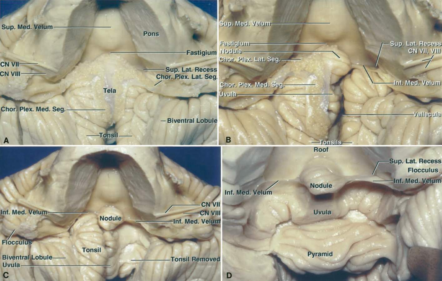
Anterior Views of the Roof of the Fourth Ventricle
7014
Surgical Correlation
Tags
Anterior Views of the Roof of the Fourth Ventricle. A, A portion of the pons and the whole medulla have been removed to provide this view into the roof of the fourth ventricle. The medial portion of the upper half of the roof is formed by the superior medullary velum. The lateral portions of the upper half of the roof, which are formed by the superior cerebellar peduncles, are hidden by the remaining pons. The fastigium is located at the junction of the upper and lower parts of the roof. The tela choroidea is the site of attachment of paired L-shaped fringes of choroid plexus, which contain the medial and lateral segments. The paired medial segments are oriented longitudinally and project from the foramen of Magendie. The lateral segments are oriented transversely and project from the foramina of Luschka. The inferior medullary velum is hidden deep with respect to the choroid plexus in this view. B, The right half of the tela choroidea has been removed to expose the nodule and the right half of the inferior medullary velum, which sweeps laterally from the surface of the nodule and blends into the flocculus at the level of the lateral recess. The superolateral recess is situated above the lateral portion of the inferior medullary velum. C, The tela choroidea on both sides and the right tonsil have been removed. The rostral pole of the tonsil bulges upward into the inferior medullary velum, but is separated from it by a narrow extension of the cerebellopontine fissure in which the PICA courses. D, Both tonsils and the uvula have been removed while preserving the inferior medullary velum. The uvula was removed to demonstrate that the inferior medullary velum connects the nodule and flocculus. (Images courtesy of AL Rhoton, Jr.)


