Anatomic Dissections of Supratentorial Cisterns
6902
Surgical Correlation
Tags
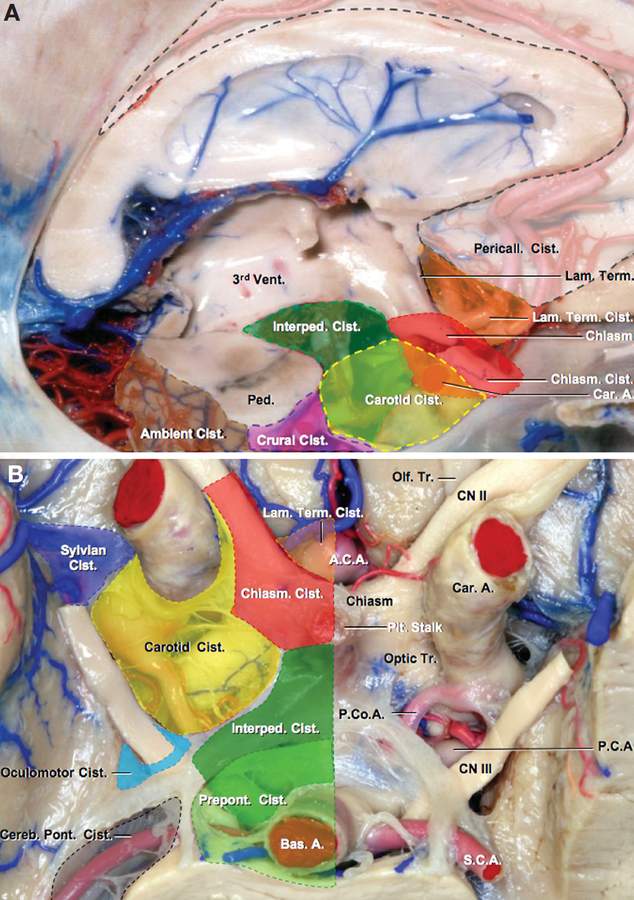
Anatomic Dissections of Supratentorial Cisterns. A, Lateral view of a midsagittal section of the brain showing the supratentorial cisterns. B, Inferior view of the basal cisterns. A and B, The supratentorial cisterns defined in this study are the pericallosal (white); lamina terminalis (orange); chiasmatic (red); interpeduncular (dark green); carotid (yellow); crural (pink); ambient (brown); sylvian (purple); and the oculomotor (light blue). The prepontine (light green) and cerebellopontine (gray) cisterns are posterior fossa cisterns. The pericallosal cistern is situated in the interhemispheric fissure on the outer surface of the corpus callosum and contains the anterior cerebral arteries. The lamina terminalis cistern is positioned above the optic chiasm and anterior to the lamina terminalis between the frontal lobes and contains the A1 and proximal A2 segments of the anterior cerebral artery, the anterior communicating and orbitofrontal arteries, some of the basal perforating arteries, and the initial segment of the recurrent artery. The chiasmatic cistern surrounds the optic nerves and chiasm and contains the pituitary stalk and carotid perforating branches. The interpeduncular cistern is situated between the cerebral peduncles and the leaves of Liliequist’s membrane at the confluence of the supra- and infratentorial parts of the subarachnoid space and contains the basilar bifurcation, proximal part of the posterior cerebral arteries, thalamoperforating arteries, and the initial segment of the circumflex perforating and medial posterior choroidal arteries. The carotid cistern is situated between the lateral edge of the optic chiasm medially, the temporal lobes laterally, and the anterior perforated substance above and contains the internal carotid and posterior communicating arteries and the origin of the anterior and middle cerebral, ophthalmic, and anterior choroidal arteries. The sylvian cistern is situated between the frontal and parietal lobes above and the temporal lobes below and contains the M1, M2, and M3 segments of the middle cerebral artery. The oculomotor cistern is formed by the confluence of the inner and the outer arachnoid membranes that converge to surround the oculomotor nerve. The crural cistern is located between the cerebral peduncle and posterior part of the uncus. The ambient cistern is positioned posterior to the crural cistern between the midbrain and the part of the medial temporal lobe behind the uncus. The crural and ambient cisterns contain the P2 segment of the posterior cerebral artery and some of the perforating, cortical, and choroidal branches arising from the P1 and P2 segments. (Images courtesy of AL Rhoton, Jr.)


